Sunday Poster Session
Category: Colon
P0240 - An Exceedingly Rare Case of Colonic Metastasis From Sinonasal Squamous Cell Carcinoma
Sunday, October 22, 2023
3:30 PM - 7:00 PM PT
Location: Exhibit Hall

Has Audio
- TG
Tyler Grantham, MD
Staten Island University Hospital
Staten Island, NY
Presenting Author(s)
Rajarajeshwari Ramachandran, MD1, Tyler Grantham, MD2, Vikash Kumar, MD1, Heidi Budke, MD3, Vinaya Gaduputi, MD3
1Brooklyn Hospital Center, Brooklyn, NY; 2Staten Island University Hospital, Staten Island, NY; 3Blanchard Valley Health System, Findlay, OH
Introduction: Sinonasal squamous cell carcinoma is a rare malignancy arising from the nasal cavity or paranasal sinuses. The incidence has been slowly declining and accounts for only 3% of head and neck cancers. Although it typically spreads locally, metastasis to the lungs, liver, and bone have been reported. Metastasis to the colon is exceedingly rare, with a limited number of cases reported in the literature. In this case report, we describe a patient who presented with severe iron deficiency anemia secondary to cecal metastasis from sinonasal squamous cell carcinoma.
Case Description/Methods: A 49-year-old man with sinonasal squamous cell carcinoma (and nodal metastasis) presented for evaluation of severe iron deficiency anemia. His hemoglobin dropped from 14 g/dL to 6.8 g/dL over 2 weeks. He endorsed exertional shortness of breath for a week prior to presentation. He denied dysphagia, nausea, vomiting, hematemesis, abdominal pain, recent change in bowel habits, melena, and hematochezia. He had no palpable abdominal mass or signs of overt gastrointestinal bleeding.
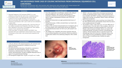
Given the symptomatic iron deficiency anemia in the setting of advanced malignancy, risks, benefits, and alternatives to endoscopic evaluation were discussed with the patient and he opted to undergo esophagogastroduodenoscopy (EGD) and colonoscopy. EGD did not demonstrate any upper gastrointestinal source of bleeding. During colonoscopy, a friable, ulcerated lesion was found in the cecum (image A). Biopsy of the cecal lesion demonstrated nests of malignant cells in the lamina propria with central keratin and necrosis (image B). Immunohistochemical stains for CK5/6, p16, p40, and p63 were positive. The findings were compatible with colonic metastasis from the known sinonasal squamous cell carcinoma. Patient succumbed to complications related to the underlying malignancy three months later.
Discussion: Risk factors of sinonasal squamous cell carcinoma include tobacco smoke, human papillomavirus infection, air pollution, occupational exposures to leather and wood dust. It is accompanied by poor 5-year overall survival of about 50%, but when patients present with metastatic disease the median survival is around 9 months. Our patient presented with severe, symptomatic iron deficiency anemia secondary to blood loss from the friable cecal metastatic lesion in the setting of primary sinonasal squamous cell carcinoma.

Disclosures:
Rajarajeshwari Ramachandran, MD1, Tyler Grantham, MD2, Vikash Kumar, MD1, Heidi Budke, MD3, Vinaya Gaduputi, MD3. P0240 - An Exceedingly Rare Case of Colonic Metastasis From Sinonasal Squamous Cell Carcinoma, ACG 2023 Annual Scientific Meeting Abstracts. Vancouver, BC, Canada: American College of Gastroenterology.
1Brooklyn Hospital Center, Brooklyn, NY; 2Staten Island University Hospital, Staten Island, NY; 3Blanchard Valley Health System, Findlay, OH
Introduction: Sinonasal squamous cell carcinoma is a rare malignancy arising from the nasal cavity or paranasal sinuses. The incidence has been slowly declining and accounts for only 3% of head and neck cancers. Although it typically spreads locally, metastasis to the lungs, liver, and bone have been reported. Metastasis to the colon is exceedingly rare, with a limited number of cases reported in the literature. In this case report, we describe a patient who presented with severe iron deficiency anemia secondary to cecal metastasis from sinonasal squamous cell carcinoma.
Case Description/Methods: A 49-year-old man with sinonasal squamous cell carcinoma (and nodal metastasis) presented for evaluation of severe iron deficiency anemia. His hemoglobin dropped from 14 g/dL to 6.8 g/dL over 2 weeks. He endorsed exertional shortness of breath for a week prior to presentation. He denied dysphagia, nausea, vomiting, hematemesis, abdominal pain, recent change in bowel habits, melena, and hematochezia. He had no palpable abdominal mass or signs of overt gastrointestinal bleeding.
Given the symptomatic iron deficiency anemia in the setting of advanced malignancy, risks, benefits, and alternatives to endoscopic evaluation were discussed with the patient and he opted to undergo esophagogastroduodenoscopy (EGD) and colonoscopy. EGD did not demonstrate any upper gastrointestinal source of bleeding. During colonoscopy, a friable, ulcerated lesion was found in the cecum (image A). Biopsy of the cecal lesion demonstrated nests of malignant cells in the lamina propria with central keratin and necrosis (image B). Immunohistochemical stains for CK5/6, p16, p40, and p63 were positive. The findings were compatible with colonic metastasis from the known sinonasal squamous cell carcinoma. Patient succumbed to complications related to the underlying malignancy three months later.
Discussion: Risk factors of sinonasal squamous cell carcinoma include tobacco smoke, human papillomavirus infection, air pollution, occupational exposures to leather and wood dust. It is accompanied by poor 5-year overall survival of about 50%, but when patients present with metastatic disease the median survival is around 9 months. Our patient presented with severe, symptomatic iron deficiency anemia secondary to blood loss from the friable cecal metastatic lesion in the setting of primary sinonasal squamous cell carcinoma.

Figure: Image A: Colonoscopy image - ulcerated cecal lesion
Image B: Histopathology image - squamous carcinoma cells in between colonic glands (black arrow)
Image B: Histopathology image - squamous carcinoma cells in between colonic glands (black arrow)
Disclosures:
Rajarajeshwari Ramachandran indicated no relevant financial relationships.
Tyler Grantham indicated no relevant financial relationships.
Vikash Kumar indicated no relevant financial relationships.
Heidi Budke indicated no relevant financial relationships.
Vinaya Gaduputi indicated no relevant financial relationships.
Rajarajeshwari Ramachandran, MD1, Tyler Grantham, MD2, Vikash Kumar, MD1, Heidi Budke, MD3, Vinaya Gaduputi, MD3. P0240 - An Exceedingly Rare Case of Colonic Metastasis From Sinonasal Squamous Cell Carcinoma, ACG 2023 Annual Scientific Meeting Abstracts. Vancouver, BC, Canada: American College of Gastroenterology.
