Sunday Poster Session
Category: Colon
P0241 - An Exceedingly Rare Case of Idiopathic Isolated Pelvic Varices
Sunday, October 22, 2023
3:30 PM - 7:00 PM PT
Location: Exhibit Hall

Has Audio
- TG
Tyler Grantham, MD
Staten Island University Hospital
Staten Island, NY
Presenting Author(s)
Tyler Grantham, MD1, Rajarajeshwari Ramachandran, MD2, Vikash Kumar, MD2, Vinaya Gaduputi, MD3
1Staten Island University Hospital, Staten Island, NY; 2Brooklyn Hospital Center, Brooklyn, NY; 3Blanchard Valley Health System, Findlay, OH
Introduction: Pelvic varices are typically associated with evidence of portal hypertension. Portal hypertension is commonly caused by liver cirrhosis, but it can also arise due to a myriad of non-cirrhotic causes. Isolated pelvic varices in the absence of portal hypertension is exceedingly rare and represents a diagnostic challenge as it can mimic other conditions, including pelvic congestion syndrome, or pelvic masses.
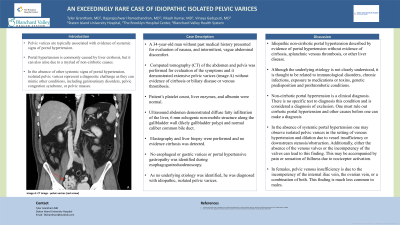
Case Description/Methods: A 34-year-old man without past medical history presented for evaluation of nausea, and intermittent, vague abdominal discomfort. Computed tomography (CT) of the abdomen and pelvis was performed for evaluation of the symptoms and it demonstrated extensive pelvic varices (image A) without evidence of cirrhosis or biliary disease or venous thrombosis. Patient’s platelet count, liver enzymes, and albumin were normal. Ultrasound abdomen demonstrated diffuse fatty infiltration of the liver, 6 mm echogenic non-mobile structure along the gallbladder wall (likely gallbladder polyp) and normal caliber common bile duct. Elastography and liver biopsy were performed and no cirrhosis was detected. No esophageal or gastric varices or portal hypertensive gastropathy was identified during esophagogastroduodenoscopy. As no underlying etiology was identified, he was diagnosed with idiopathic, isolated pelvic varices.
Discussion: In the absence of portal hypertension, isolated pelvic varices can be seen in the setting of venous obstruction, which may be accompanied by pain or sensation of fullness due to nociceptor activation. Imaging performed in our patient did not demonstrate venous obstruction leading to the diagnosis of idiopathic, isolated pelvic varices.

Disclosures:
Tyler Grantham, MD1, Rajarajeshwari Ramachandran, MD2, Vikash Kumar, MD2, Vinaya Gaduputi, MD3. P0241 - An Exceedingly Rare Case of Idiopathic Isolated Pelvic Varices, ACG 2023 Annual Scientific Meeting Abstracts. Vancouver, BC, Canada: American College of Gastroenterology.
1Staten Island University Hospital, Staten Island, NY; 2Brooklyn Hospital Center, Brooklyn, NY; 3Blanchard Valley Health System, Findlay, OH
Introduction: Pelvic varices are typically associated with evidence of portal hypertension. Portal hypertension is commonly caused by liver cirrhosis, but it can also arise due to a myriad of non-cirrhotic causes. Isolated pelvic varices in the absence of portal hypertension is exceedingly rare and represents a diagnostic challenge as it can mimic other conditions, including pelvic congestion syndrome, or pelvic masses.
Case Description/Methods: A 34-year-old man without past medical history presented for evaluation of nausea, and intermittent, vague abdominal discomfort. Computed tomography (CT) of the abdomen and pelvis was performed for evaluation of the symptoms and it demonstrated extensive pelvic varices (image A) without evidence of cirrhosis or biliary disease or venous thrombosis. Patient’s platelet count, liver enzymes, and albumin were normal. Ultrasound abdomen demonstrated diffuse fatty infiltration of the liver, 6 mm echogenic non-mobile structure along the gallbladder wall (likely gallbladder polyp) and normal caliber common bile duct. Elastography and liver biopsy were performed and no cirrhosis was detected. No esophageal or gastric varices or portal hypertensive gastropathy was identified during esophagogastroduodenoscopy. As no underlying etiology was identified, he was diagnosed with idiopathic, isolated pelvic varices.
Discussion: In the absence of portal hypertension, isolated pelvic varices can be seen in the setting of venous obstruction, which may be accompanied by pain or sensation of fullness due to nociceptor activation. Imaging performed in our patient did not demonstrate venous obstruction leading to the diagnosis of idiopathic, isolated pelvic varices.

Figure: Image A: CT image - pelvic varices (red arrow)
Disclosures:
Tyler Grantham indicated no relevant financial relationships.
Rajarajeshwari Ramachandran indicated no relevant financial relationships.
Vikash Kumar indicated no relevant financial relationships.
Vinaya Gaduputi indicated no relevant financial relationships.
Tyler Grantham, MD1, Rajarajeshwari Ramachandran, MD2, Vikash Kumar, MD2, Vinaya Gaduputi, MD3. P0241 - An Exceedingly Rare Case of Idiopathic Isolated Pelvic Varices, ACG 2023 Annual Scientific Meeting Abstracts. Vancouver, BC, Canada: American College of Gastroenterology.
