Sunday Poster Session
Category: Colon
P0244 - Cap Polyposis Syndrome Imitating Inflammatory Bowel Disease With Polyps Extending to the Terminal Ileum
Sunday, October 22, 2023
3:30 PM - 7:00 PM PT
Location: Exhibit Hall

Has Audio

Garrett T. Coleman, BBA
UTMB
Galveston, TX
Presenting Author(s)
Garrett T. Coleman, BBA1, Rawan Dayah, MD2, Suimin Qiu, MD, PhD1, Gurinder Luthra, DO1
1UTMB, Galveston, TX; 2UTMB, Webster, TX
Introduction: Cap polyposis syndrome is a rare condition characterized by erythematous, inflammatory colonic polyps covered by a cap of fibrinopurulent mucous. We describe the case of a 34-year-old male who presented with diarrhea and history of hundreds of colonic polyps. While most cases of cap polyposis are confined to the distal colon and rectum, we believe this is the first documented case of cap polyposis extending to the terminal ileum.
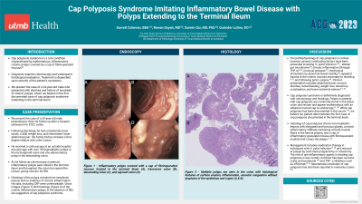
Case Description/Methods: We present the case of a 34-year-old male presenting to clinic for follow up after a hospital admission for ETEC colitis. Following discharge, he had consistently loose stools, a 40lb weight loss, and intermittent lower abdominal pain that ultimately resolved. The patient has a family history significant for colon cancer and received a colonoscopy at an outside hospital one year ago with over 100 hyperplastic polyps in the rectosigmoid colon and one adenomatous polyp in the descending colon. At his follow-up colonoscopy, numerous polyps were noted in the terminal ileum and throughout the colon. Endoscopically, the polyps appeared similar to pseudopolyps, giving concern for the presence of Inflammatory Bowel Disease (IBD). Several cold snare polypectomies were performed throughout the colon revealing inflammatory polyps. The patient has no evidence of chronic inflammation or IBD. Gross images of the colonic inflammatory polyps (Figures C-F) were noted to have a cap of fibrinopurulent mucous. Histology of the inflammatory polyps (Figures A-B) demonstrated surface erosion, inflammation, and vascular congestion without dysplasia of the epithelium or crypts. The endoscopic and histologic features in the absence of IBD are suggestive of cap polyposis.
Discussion: The clinical presentation of cap polyposis syndrome is nonspecific and may mimic conditions such as IBD or colorectal cancer. Patients most commonly present with abdominal pain, mucoid diarrhea, and rectal bleeding. Patients may also present with weight loss, tenesmus, and constipation. Laboratory values may reveal iron deficiency anemia, hypoproteinemia, and normal C-reactive protein. Cap polyposis is definitively diagnosed with erythematous polyps with an adherent mucoid cap on endoscopy and non-neoplastic lesions with elongated and tortuous glands, a mixed inflammatory infiltrate containing smooth muscle fibers in the lamina propria, and a cap of inflammatory granulation tissue with fibrinopurulent exudate that covers the polyps on histology.

Disclosures:
Garrett T. Coleman, BBA1, Rawan Dayah, MD2, Suimin Qiu, MD, PhD1, Gurinder Luthra, DO1. P0244 - Cap Polyposis Syndrome Imitating Inflammatory Bowel Disease With Polyps Extending to the Terminal Ileum, ACG 2023 Annual Scientific Meeting Abstracts. Vancouver, BC, Canada: American College of Gastroenterology.
1UTMB, Galveston, TX; 2UTMB, Webster, TX
Introduction: Cap polyposis syndrome is a rare condition characterized by erythematous, inflammatory colonic polyps covered by a cap of fibrinopurulent mucous. We describe the case of a 34-year-old male who presented with diarrhea and history of hundreds of colonic polyps. While most cases of cap polyposis are confined to the distal colon and rectum, we believe this is the first documented case of cap polyposis extending to the terminal ileum.
Case Description/Methods: We present the case of a 34-year-old male presenting to clinic for follow up after a hospital admission for ETEC colitis. Following discharge, he had consistently loose stools, a 40lb weight loss, and intermittent lower abdominal pain that ultimately resolved. The patient has a family history significant for colon cancer and received a colonoscopy at an outside hospital one year ago with over 100 hyperplastic polyps in the rectosigmoid colon and one adenomatous polyp in the descending colon. At his follow-up colonoscopy, numerous polyps were noted in the terminal ileum and throughout the colon. Endoscopically, the polyps appeared similar to pseudopolyps, giving concern for the presence of Inflammatory Bowel Disease (IBD). Several cold snare polypectomies were performed throughout the colon revealing inflammatory polyps. The patient has no evidence of chronic inflammation or IBD. Gross images of the colonic inflammatory polyps (Figures C-F) were noted to have a cap of fibrinopurulent mucous. Histology of the inflammatory polyps (Figures A-B) demonstrated surface erosion, inflammation, and vascular congestion without dysplasia of the epithelium or crypts. The endoscopic and histologic features in the absence of IBD are suggestive of cap polyposis.
Discussion: The clinical presentation of cap polyposis syndrome is nonspecific and may mimic conditions such as IBD or colorectal cancer. Patients most commonly present with abdominal pain, mucoid diarrhea, and rectal bleeding. Patients may also present with weight loss, tenesmus, and constipation. Laboratory values may reveal iron deficiency anemia, hypoproteinemia, and normal C-reactive protein. Cap polyposis is definitively diagnosed with erythematous polyps with an adherent mucoid cap on endoscopy and non-neoplastic lesions with elongated and tortuous glands, a mixed inflammatory infiltrate containing smooth muscle fibers in the lamina propria, and a cap of inflammatory granulation tissue with fibrinopurulent exudate that covers the polyps on histology.

Figure: Multiple polyps are seen in the colon with histological features of surface erosion, inflammation, vascular congestions without dysplasia of the epithelium and crypts (A & B). Inflammatory polyps covered with a cap of fibrinopurulent mucous located in the terminal ileum (C), transverse colon (D), descending colon (E), and sigmoid colon (F).
Disclosures:
Garrett Coleman indicated no relevant financial relationships.
Rawan Dayah indicated no relevant financial relationships.
Suimin Qiu indicated no relevant financial relationships.
Gurinder Luthra indicated no relevant financial relationships.
Garrett T. Coleman, BBA1, Rawan Dayah, MD2, Suimin Qiu, MD, PhD1, Gurinder Luthra, DO1. P0244 - Cap Polyposis Syndrome Imitating Inflammatory Bowel Disease With Polyps Extending to the Terminal Ileum, ACG 2023 Annual Scientific Meeting Abstracts. Vancouver, BC, Canada: American College of Gastroenterology.

