Sunday Poster Session
Category: Colon
P0246 - Plug-Assisted Retrograde Transvenous Obliteration (PARTO) for Cecal Varices Treatment
Sunday, October 22, 2023
3:30 PM - 7:00 PM PT
Location: Exhibit Hall

Has Audio

Sherry Dehghani, MD
Montefiore Medical Center
New York, NY
Presenting Author(s)
Sherry Dehghani, MD, Christopher Andrade, MD, Hilary Hertan, MD
Montefiore Medical Center, New York, NY
Introduction: Bleeding varices in cirrhosis are primarily found in the distal esophagus.1 However, ectopic varices can develop in non-esophageal or gastric areas. Among these, colonic varices, especially cecal varices, are rare.2 Here, we present a case of life-threatening cecal variceal hemorrhage successfully treated with endoscopic and vascular interventions.
Case Description/Methods: The patient, a 71-year-old male with a medical history of alcohol-related cirrhosis, presented with one-day hematochezia. He had complications including esophageal varices, hepatic encephalopathy, and hepatocellular carcinoma. Upon admission, the patient was hypotensive and anemic, necessitating massive transfusion and vasopressor support in the ICU. During upper endoscopy, recent bleeding was observed in two small varices, which were successfully treated with banding. Colonoscopy revealed active bleeding from a visible vessel at the appendiceal orifice, stopped by the placement of three hemostatic clips. CT angiogram indicated no acute GI bleeding, but intervention radiology (IR) was consulted for embolization. The angiogram revealed a large tortuous portosystemic varix with inflow from the superior mesenteric vein and outflow through the right gonadal vein and a smaller branch to the inferior vena cava. The patient underwent Plug-assisted retrograde transvenous obliteration (PARTO) of the cecal varix using a 1.3cm Microvascular Plug (MVP). Post-procedure angiography confirmed successful obliteration of the varix. Unfortunately, the patient passed away due to septic shock caused by pneumonia.
Discussion: Portal hypertension causes esophageal and extraesophageal varices, primarily in the stomach and rectum. Although colonic varices are rare, identifying them is crucial due to potential severe hemorrhage.3 Colonoscopy is the gold standard for diagnosing cecal varices, but caution is necessary to avoid variceal collapse due to over-insufflation or hypotension. CT angiography offers a minimally invasive option for localizing colonic varices.2 There are no established guidelines for treating extraesophageal varices, but endoscopic interventions are used for unstable patients ineligible for TIPS, liver transplantation, or colectomy. Sclerotherapy and band ligation show varying levels of safety and efficacy.2 BRTO and PARTO are commonly used for gastric varices but rarely for cecal varices.4 This case contributes valuable data to the literature, emphasizing the need for long-term follow-up to assess efficacy.

Disclosures:
Sherry Dehghani, MD, Christopher Andrade, MD, Hilary Hertan, MD. P0246 - Plug-Assisted Retrograde Transvenous Obliteration (PARTO) for Cecal Varices Treatment, ACG 2023 Annual Scientific Meeting Abstracts. Vancouver, BC, Canada: American College of Gastroenterology.
Montefiore Medical Center, New York, NY
Introduction: Bleeding varices in cirrhosis are primarily found in the distal esophagus.1 However, ectopic varices can develop in non-esophageal or gastric areas. Among these, colonic varices, especially cecal varices, are rare.2 Here, we present a case of life-threatening cecal variceal hemorrhage successfully treated with endoscopic and vascular interventions.
Case Description/Methods: The patient, a 71-year-old male with a medical history of alcohol-related cirrhosis, presented with one-day hematochezia. He had complications including esophageal varices, hepatic encephalopathy, and hepatocellular carcinoma. Upon admission, the patient was hypotensive and anemic, necessitating massive transfusion and vasopressor support in the ICU. During upper endoscopy, recent bleeding was observed in two small varices, which were successfully treated with banding. Colonoscopy revealed active bleeding from a visible vessel at the appendiceal orifice, stopped by the placement of three hemostatic clips. CT angiogram indicated no acute GI bleeding, but intervention radiology (IR) was consulted for embolization. The angiogram revealed a large tortuous portosystemic varix with inflow from the superior mesenteric vein and outflow through the right gonadal vein and a smaller branch to the inferior vena cava. The patient underwent Plug-assisted retrograde transvenous obliteration (PARTO) of the cecal varix using a 1.3cm Microvascular Plug (MVP). Post-procedure angiography confirmed successful obliteration of the varix. Unfortunately, the patient passed away due to septic shock caused by pneumonia.
Discussion: Portal hypertension causes esophageal and extraesophageal varices, primarily in the stomach and rectum. Although colonic varices are rare, identifying them is crucial due to potential severe hemorrhage.3 Colonoscopy is the gold standard for diagnosing cecal varices, but caution is necessary to avoid variceal collapse due to over-insufflation or hypotension. CT angiography offers a minimally invasive option for localizing colonic varices.2 There are no established guidelines for treating extraesophageal varices, but endoscopic interventions are used for unstable patients ineligible for TIPS, liver transplantation, or colectomy. Sclerotherapy and band ligation show varying levels of safety and efficacy.2 BRTO and PARTO are commonly used for gastric varices but rarely for cecal varices.4 This case contributes valuable data to the literature, emphasizing the need for long-term follow-up to assess efficacy.

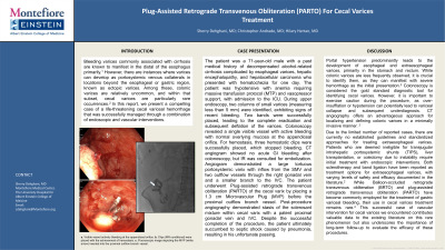
Figure: a. Visible vessel actively bleeding at the appendiceal orifice. b. Clips (MR conditional) were placed with the achievement of hemostasis. c. Fluoroscopic image depicting the MVP (white arrow) inserted into the proximal outflow branch vessel.
Disclosures:
Sherry Dehghani indicated no relevant financial relationships.
Christopher Andrade indicated no relevant financial relationships.
Hilary Hertan indicated no relevant financial relationships.
Sherry Dehghani, MD, Christopher Andrade, MD, Hilary Hertan, MD. P0246 - Plug-Assisted Retrograde Transvenous Obliteration (PARTO) for Cecal Varices Treatment, ACG 2023 Annual Scientific Meeting Abstracts. Vancouver, BC, Canada: American College of Gastroenterology.
