Sunday Poster Session
Category: Colon
P0252 - A Twisted Tale: Sigmoid Volvulus Secondary to Rectal Stricture
Sunday, October 22, 2023
3:30 PM - 7:00 PM PT
Location: Exhibit Hall

Has Audio

Suvithan Rajadurai, MD
Warren Alpert Medical School of Brown University
Providence, RI
Presenting Author(s)
Suvithan Rajadurai, MD1, Andrew Krane, MD2, Colleen R. Kelly, MD, FACG3, Kaio Ferreira, MD4
1Warren Alpert Medical School of Brown University, Providence, RI; 2Warren Alpert Medical School, Brown University, Providence, RI; 3Alpert School of Medicine of Brown University, Providence, RI; 4Brown University, Providence, RI
Introduction: Endometriosis describes a condition in which endometrial glands and stroma occur outside the normal uterine cavity. Lesions are typically localized to the pelvis but can present to extrapelvic locations such as the bowel, rarely manifesting as stricture. Here we present a case of sigmoid volvulus possibly secondary to chronic endometriotic stricture
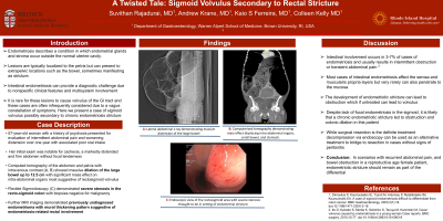
Case Description/Methods: A 57-year-old woman with a history of mood disorder and psychosis presented for evaluation of one year of intermittent abdominal pain and worsening distension with associated poor oral intake. Her initial exam was notable for cachexia, and a markedly distended, firm abdomen without focal tenderness. Labs were notable for potassium of 2.6 mEq/L, sodium of 146 mmol/L, white blood cell count 9.2x10⁹/L, albumin 2.2 u/L, and normal liver enzymes. Lactate was within normal limits. Computed tomography of the abdomen and pelvis with intravenous contrast (Image 1) showed massive dilation of the large bowel up to 12.5 cm with mass effect on intra-abdominal organs suggestive of rectosigmoid volvulus alongside a focal area of wall thickening. A flexible sigmoidoscopy was performed which showed an obstructing stenotic area that was unable to be traversed. Colorectal surgery was consulted for exploratory laparotomy; total abdominal colectomy with ileostomy and rectosigmoid fistula was performed. Biopsies were obtained of the colon, appendix, terminal ileum and lymph nodes. Gross colon specimen showed no mass, appendiceal biopsy showed prominent endometriosis with serosal adhesions. Lymph nodes were negative for malignancy. A repeat sigmoidoscopy was performed to obtain additional biopsies of the rectal stricture, which showed marked acute inflammation and granulation tissue. Pelvic MRI showed a right ovarian lesion most suggestive of an endometrioma. The case was discussed at tumor board and it was felt the thickening was secondary to chronic stricture although etiology of the stricture remains inconclusive at this time.
Discussion: Endometriotic stricture leading to obstruction is rare but if untreated can lead to volvulus. Despite lack of focal endometriosis in the sigmoid, it is possible that a chronic endometriotic stricture led to obstruction and colonic dilation In scenarios with recurrent abdominal pain, and bowel obstruction in a reproductive age female patient, endometriosis should be in the differential diagnosis.

Disclosures:
Suvithan Rajadurai, MD1, Andrew Krane, MD2, Colleen R. Kelly, MD, FACG3, Kaio Ferreira, MD4. P0252 - A Twisted Tale: Sigmoid Volvulus Secondary to Rectal Stricture, ACG 2023 Annual Scientific Meeting Abstracts. Vancouver, BC, Canada: American College of Gastroenterology.
1Warren Alpert Medical School of Brown University, Providence, RI; 2Warren Alpert Medical School, Brown University, Providence, RI; 3Alpert School of Medicine of Brown University, Providence, RI; 4Brown University, Providence, RI
Introduction: Endometriosis describes a condition in which endometrial glands and stroma occur outside the normal uterine cavity. Lesions are typically localized to the pelvis but can present to extrapelvic locations such as the bowel, rarely manifesting as stricture. Here we present a case of sigmoid volvulus possibly secondary to chronic endometriotic stricture
Case Description/Methods: A 57-year-old woman with a history of mood disorder and psychosis presented for evaluation of one year of intermittent abdominal pain and worsening distension with associated poor oral intake. Her initial exam was notable for cachexia, and a markedly distended, firm abdomen without focal tenderness. Labs were notable for potassium of 2.6 mEq/L, sodium of 146 mmol/L, white blood cell count 9.2x10⁹/L, albumin 2.2 u/L, and normal liver enzymes. Lactate was within normal limits. Computed tomography of the abdomen and pelvis with intravenous contrast (Image 1) showed massive dilation of the large bowel up to 12.5 cm with mass effect on intra-abdominal organs suggestive of rectosigmoid volvulus alongside a focal area of wall thickening. A flexible sigmoidoscopy was performed which showed an obstructing stenotic area that was unable to be traversed. Colorectal surgery was consulted for exploratory laparotomy; total abdominal colectomy with ileostomy and rectosigmoid fistula was performed. Biopsies were obtained of the colon, appendix, terminal ileum and lymph nodes. Gross colon specimen showed no mass, appendiceal biopsy showed prominent endometriosis with serosal adhesions. Lymph nodes were negative for malignancy. A repeat sigmoidoscopy was performed to obtain additional biopsies of the rectal stricture, which showed marked acute inflammation and granulation tissue. Pelvic MRI showed a right ovarian lesion most suggestive of an endometrioma. The case was discussed at tumor board and it was felt the thickening was secondary to chronic stricture although etiology of the stricture remains inconclusive at this time.
Discussion: Endometriotic stricture leading to obstruction is rare but if untreated can lead to volvulus. Despite lack of focal endometriosis in the sigmoid, it is possible that a chronic endometriotic stricture led to obstruction and colonic dilation In scenarios with recurrent abdominal pain, and bowel obstruction in a reproductive age female patient, endometriosis should be in the differential diagnosis.

Figure: Figure 1. (A): Lateral CT view of abdomen, (B) Anterior Posterior view of CT abdomen, note the multiple distended loops of bowel. (C) Coronal CT view of abdomen redemonstrating distension (D) Lateral CT view
Disclosures:
Suvithan Rajadurai indicated no relevant financial relationships.
Andrew Krane indicated no relevant financial relationships.
Colleen Kelly: Sebela Pharmaceuticals – Consultant.
Kaio Ferreira indicated no relevant financial relationships.
Suvithan Rajadurai, MD1, Andrew Krane, MD2, Colleen R. Kelly, MD, FACG3, Kaio Ferreira, MD4. P0252 - A Twisted Tale: Sigmoid Volvulus Secondary to Rectal Stricture, ACG 2023 Annual Scientific Meeting Abstracts. Vancouver, BC, Canada: American College of Gastroenterology.
