Sunday Poster Session
Category: Colon
P0257 - An Unfortunate Diagnosis After Foreign Bodies Identified in the Sigmoid Colon
Sunday, October 22, 2023
3:30 PM - 7:00 PM PT
Location: Exhibit Hall

Has Audio

Jeremy Polman, DO, MS, MBA
St. David's South Austin Medical Center
New York, NY
Presenting Author(s)
Jeremy Polman, DO, MS, MBA1, Lauren Lyssy, DO2
1St. David's South Austin Medical Center, Nashville, TN; 2St. David's South Austin Medical Center, Austin, TX
Introduction: Foreign body ingestion can often cause bowel obstruction. Rarely, an ingested foreign body can unintentionally aid in the diagnosis of further pathology.
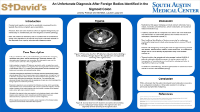
Case Description/Methods: A 33-year-old female with no past medical history presented to the emergency department with crampy, diffuse abdominal pain, abdominal distention, and decreasing stool frequency over two weeks. She attempted to use over-the-counter calcium carbonate without significant relief. Initial evaluation included a CT abdomen and pelvis, which revealed two round, radiopaque foreign objects in the sigmoid colon and segmental luminal narrowing of the sigmoid colon. Imaging also demonstrated multiple hypodense liver lesions and a complex right adnexal/ovarian lesion. Gastroenterology and general surgery were then consulted, and a multi-specialty discussion was held to decide the best therapeutic approach. A flexible sigmoidoscopy performed the following morning demonstrated severe stenosis about 20cm from the anal verge, which measured 4cm in length by 7mm in diameter. Inspection of the stenotic area demonstrated what appeared to be a foreign body obstructing the opening of the stricture. The endoscope was advanced through the stricture with the assistance of spray water, which dislodged the foreign object and washed it proximally. The colon proximal to the stricture was dilated and filled with stool, preventing further advancement of the endoscope. The foreign objects appeared significantly larger in diameter than the opening of the stenotic area, and the procedure was terminated. Exploratory laparotomy was completed the following morning, which surprisingly demonstrated severe carcinomatosis with numerous peritoneal implants, omental caking, invasion of the cystic/common bile duct junction, invasion of the sigmoid colon, and a right ovary covered in tumor. Ultimately, the sigmoid colon was resected, end colostomy placed, and multiple biopsies were obtained, which revealed poorly differentiated mucinous adenocarcinoma, suspicious for mucinous ovarian malignancy. While originally presumed to be metallic, gross inspection of the foreign bodies appeared to demonstrate medication filled capsules.
Discussion: Medications that contain calcium carbonate, iron, phenothiazines or those with enteric coating may appear radiopaque on imaging. While unfortunate that the patient developed bowel obstruction secondary to an undigested medication, it fortunately assisted in revealing a previously asymptomatic malignancy.

Disclosures:
Jeremy Polman, DO, MS, MBA1, Lauren Lyssy, DO2. P0257 - An Unfortunate Diagnosis After Foreign Bodies Identified in the Sigmoid Colon, ACG 2023 Annual Scientific Meeting Abstracts. Vancouver, BC, Canada: American College of Gastroenterology.
1St. David's South Austin Medical Center, Nashville, TN; 2St. David's South Austin Medical Center, Austin, TX
Introduction: Foreign body ingestion can often cause bowel obstruction. Rarely, an ingested foreign body can unintentionally aid in the diagnosis of further pathology.
Case Description/Methods: A 33-year-old female with no past medical history presented to the emergency department with crampy, diffuse abdominal pain, abdominal distention, and decreasing stool frequency over two weeks. She attempted to use over-the-counter calcium carbonate without significant relief. Initial evaluation included a CT abdomen and pelvis, which revealed two round, radiopaque foreign objects in the sigmoid colon and segmental luminal narrowing of the sigmoid colon. Imaging also demonstrated multiple hypodense liver lesions and a complex right adnexal/ovarian lesion. Gastroenterology and general surgery were then consulted, and a multi-specialty discussion was held to decide the best therapeutic approach. A flexible sigmoidoscopy performed the following morning demonstrated severe stenosis about 20cm from the anal verge, which measured 4cm in length by 7mm in diameter. Inspection of the stenotic area demonstrated what appeared to be a foreign body obstructing the opening of the stricture. The endoscope was advanced through the stricture with the assistance of spray water, which dislodged the foreign object and washed it proximally. The colon proximal to the stricture was dilated and filled with stool, preventing further advancement of the endoscope. The foreign objects appeared significantly larger in diameter than the opening of the stenotic area, and the procedure was terminated. Exploratory laparotomy was completed the following morning, which surprisingly demonstrated severe carcinomatosis with numerous peritoneal implants, omental caking, invasion of the cystic/common bile duct junction, invasion of the sigmoid colon, and a right ovary covered in tumor. Ultimately, the sigmoid colon was resected, end colostomy placed, and multiple biopsies were obtained, which revealed poorly differentiated mucinous adenocarcinoma, suspicious for mucinous ovarian malignancy. While originally presumed to be metallic, gross inspection of the foreign bodies appeared to demonstrate medication filled capsules.
Discussion: Medications that contain calcium carbonate, iron, phenothiazines or those with enteric coating may appear radiopaque on imaging. While unfortunate that the patient developed bowel obstruction secondary to an undigested medication, it fortunately assisted in revealing a previously asymptomatic malignancy.

Figure: Figure A: Anterior-posterior slice of CT abdomen and pelvis demonstrating a round, radiopaque foreign object in the sigmoid colon and segmental luminal narrowing of the sigmoid colon.
Figure B: Coronal slice of CT abdomen and pelvis demonstrating two round, radiopaque foreign objects in the sigmoid colon and segmental luminal narrowing of the sigmoid colon.
Figure B: Coronal slice of CT abdomen and pelvis demonstrating two round, radiopaque foreign objects in the sigmoid colon and segmental luminal narrowing of the sigmoid colon.
Disclosures:
Jeremy Polman indicated no relevant financial relationships.
Lauren Lyssy indicated no relevant financial relationships.
Jeremy Polman, DO, MS, MBA1, Lauren Lyssy, DO2. P0257 - An Unfortunate Diagnosis After Foreign Bodies Identified in the Sigmoid Colon, ACG 2023 Annual Scientific Meeting Abstracts. Vancouver, BC, Canada: American College of Gastroenterology.
