Sunday Poster Session
Category: Colon
P0262 - A Rare Aggressive Colorectal Malignancy With Elevated Alpha Feto Protein
Sunday, October 22, 2023
3:30 PM - 7:00 PM PT
Location: Exhibit Hall

Has Audio

Dhanush Hoskere, DO
CarePoint Health
Bayonne, NJ
Presenting Author(s)
Dhanush Hoskere, DO1, John Hahn, MD2, Antonios Tsompanidis, DO2
1CarePoint Health, Bayonne, NJ; 2CarePoint Health - Bayonne Medical Center, Bayonne, NJ
Introduction: Colorectal adenocarcinoma with enteroblastic/yolk sac differentiation (CAED) is a rare aggressive malignancy. These tumors commonly produce alpha feto protein (AFP) and can be mistaken for hepatocellular, ovarian, or testicular carcinoma. Here, we present a rare case of right sided CAED with metastasis to the liver and gallbladder.
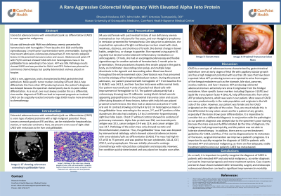
Case Description/Methods: 84 year old female with past medical history of iron deficiency anemia presented for ten episodes of hematochezia starting one day prior to admission. Of note, esophagoduodenoscopy and flexible sigmoidoscopy 1 month prior to presentation showed multiple large diverticula in the sigmoid colon without evidence of bleeding. During the most recent admission, the patient presented with hemoglobin of 7 from baseline 8 to 9 and liver function tests were within normal limits. Colonoscopy showed two 25 millimeter oozing bluish tinted necrotic appearing polypoid lesions in the proximal transverse colon and cecum. Abdomen and pelvis computed tomography with oral and intravenous contrast showed a 9.8 by 8.4 by 9.2 centimeter heterogenous mass in the gallbladder fossa extending to the cecum. AFP was 506, carcinoembryonic antigen (CEA) was 10.5, cancer antigen 19-9 (CA 19-9) was 22.4, and cancer antigen 125 (CA-125) was 14.7. Pathology of the colon mass was initially delayed due to its poor differentiation. Pathology results showed CAED and was positive for spalt like transcription factor 4 (SALL4) and glypician-3 (GP3). Patient was planned to commence chemotherapy with carboplatin and etoposide. However, she quickly deteriorated and was placed on hospice.
Discussion: CAED is rare, aggressive, and is characterized by fetal gastrointestinal epithelium. While most AFP-producing tumors are produced by the foregut endoderm, CAED originates from the hindgut endoderm. More specific tumor markers including GP3 and SALL4 help differentiate this from other AFP producing tumors. Most cases of CAED are male and the tumors generally originate in the left colon. However, our patient was female and her CAED originated in the right colon. Thus, it is imperative to consider this diagnosis with consultation of the pathologist for any patient with elevated AFP and aggressive colorectal malignancy, as our patient’s diagnosis was delayed because the specimen stained poorly due to its poor cellular differentiation. Earlier diagnosis of CAED can lead to improved prognosis, as isolated CAED can be surgically resected and early-stage CAED may be more amenable to chemotherapy.

Disclosures:
Dhanush Hoskere, DO1, John Hahn, MD2, Antonios Tsompanidis, DO2. P0262 - A Rare Aggressive Colorectal Malignancy With Elevated Alpha Feto Protein, ACG 2023 Annual Scientific Meeting Abstracts. Vancouver, BC, Canada: American College of Gastroenterology.
1CarePoint Health, Bayonne, NJ; 2CarePoint Health - Bayonne Medical Center, Bayonne, NJ
Introduction: Colorectal adenocarcinoma with enteroblastic/yolk sac differentiation (CAED) is a rare aggressive malignancy. These tumors commonly produce alpha feto protein (AFP) and can be mistaken for hepatocellular, ovarian, or testicular carcinoma. Here, we present a rare case of right sided CAED with metastasis to the liver and gallbladder.
Case Description/Methods: 84 year old female with past medical history of iron deficiency anemia presented for ten episodes of hematochezia starting one day prior to admission. Of note, esophagoduodenoscopy and flexible sigmoidoscopy 1 month prior to presentation showed multiple large diverticula in the sigmoid colon without evidence of bleeding. During the most recent admission, the patient presented with hemoglobin of 7 from baseline 8 to 9 and liver function tests were within normal limits. Colonoscopy showed two 25 millimeter oozing bluish tinted necrotic appearing polypoid lesions in the proximal transverse colon and cecum. Abdomen and pelvis computed tomography with oral and intravenous contrast showed a 9.8 by 8.4 by 9.2 centimeter heterogenous mass in the gallbladder fossa extending to the cecum. AFP was 506, carcinoembryonic antigen (CEA) was 10.5, cancer antigen 19-9 (CA 19-9) was 22.4, and cancer antigen 125 (CA-125) was 14.7. Pathology of the colon mass was initially delayed due to its poor differentiation. Pathology results showed CAED and was positive for spalt like transcription factor 4 (SALL4) and glypician-3 (GP3). Patient was planned to commence chemotherapy with carboplatin and etoposide. However, she quickly deteriorated and was placed on hospice.
Discussion: CAED is rare, aggressive, and is characterized by fetal gastrointestinal epithelium. While most AFP-producing tumors are produced by the foregut endoderm, CAED originates from the hindgut endoderm. More specific tumor markers including GP3 and SALL4 help differentiate this from other AFP producing tumors. Most cases of CAED are male and the tumors generally originate in the left colon. However, our patient was female and her CAED originated in the right colon. Thus, it is imperative to consider this diagnosis with consultation of the pathologist for any patient with elevated AFP and aggressive colorectal malignancy, as our patient’s diagnosis was delayed because the specimen stained poorly due to its poor cellular differentiation. Earlier diagnosis of CAED can lead to improved prognosis, as isolated CAED can be surgically resected and early-stage CAED may be more amenable to chemotherapy.

Figure: Top left picture is mass in proximal transverse colon. Top right picture is mass in cecum. Bottom left picture is coronal view of CT showing colon mass extending to gallbladder fossa. Bottom right picture shows histology of the colon mass, showing polygonal tumor cells with eosinophilic cytoplasm, large and hyperchromatic nuclei, arranged in thick trabeculae and solid nests with focal glandular formation
Disclosures:
Dhanush Hoskere indicated no relevant financial relationships.
John Hahn indicated no relevant financial relationships.
Antonios Tsompanidis indicated no relevant financial relationships.
Dhanush Hoskere, DO1, John Hahn, MD2, Antonios Tsompanidis, DO2. P0262 - A Rare Aggressive Colorectal Malignancy With Elevated Alpha Feto Protein, ACG 2023 Annual Scientific Meeting Abstracts. Vancouver, BC, Canada: American College of Gastroenterology.
