Sunday Poster Session
Category: Colon
P0288 - Crohn’s and Bowel Lymphoma: Non-Identical Twins?
Sunday, October 22, 2023
3:30 PM - 7:00 PM PT
Location: Exhibit Hall

Has Audio

Anhthu Tong, DO
University Hospitals
Parma, OH
Presenting Author(s)
Anhthu Tong, DO1, Vibhu Chittajallu, MD2, John Dumot, DO3, Fady G. Haddad, MD1
1University Hospitals, Parma, OH; 2Case Western Reserve University / University Hospitals, Cleveland, OH; 3University Hospitals Cleveland Medical Center, Cleveland, OH
Introduction: Gastrointestinal (GI) lymphoma constitutes 5-10% of cases of non-Hodgkin lymphoma. The most commonly affected GI sites are the stomach (60%), small intestine (20-30%) and less frequently the colon and rectum (10-20%). Despite that GI lymphoma is described in the literature, primary colon lymphoma is extremely rare comprising < 1% of all colorectal malignancies. Herein, we report the case of an adolescent boy presenting with abdominal pain secondary to colorectal lymphoma.
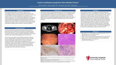
Case Description/Methods: An 18 year-old healthy boy presented to the ED for few-week history of left lower quadrant abdominal (LLQ) pain. Physical exam showed LLQ tenderness with no rebound or guarding. His twin sister and older sister are both diagnosed with Crohn’s disease. Lab tests were significant for Hb of 14.4 g/dL (normal range 13.5-17.5 g/dL), WBC of 5.9 x10E9/L (normal range 4.4-11.3 x10E9/L), lymphocyte count of 0.79 x10E9/L (normal range 1.20-4.80 x10E9/L), plts of 132 x10E9/L (normal range 150-450 x10E9/L), calcium of 11.2 mg/dL (normal range 8.6-10.6 mg/dL). CT scan of the abdomen and pelvis showed wall thickening of the rectum and sigmoid colon with extensive pelvic lymph nodes. Colonoscopy showed a large partially obstructing mass at the recto-sigmoid junction with multiple scattered polypoid lesions throughout the colon. Pathology revealed lymphoid proliferation consistent with extranodal marginal zone B-cell lymphoma (MALT). No bone marrow (BM) or distant sites involvement were noted on BM biopsy and PET scan respectively suggesting stage IIE disease. Patient was started on Rituximab with resolution of symptoms and complete response after 10 month follow-up.
Discussion: The most common primary colonic malignancies include carcinoma followed by carcinoid and GI lymphoma (< 1% of cases). The most common type of primary GI lymphoma is diffuse large B cell lymphoma, a subtype of non-Hodgkin lymphoma. The cecum is most commonly affected followed by right colon and sigmoid colon. Symptoms are nonspecific and include abdominal pain, hematochezia, and bowel obstruction, among others. Contrast abdominal CT scan suggests mucosal changes, extra-luminal extension and nodal involvement. Diagnosis relies on endoscopic visualization and tissue biopsies. The Lugano and Paris model is used for staging of gastrointestinal lymphomas. Treatment of choice is anti-CD20 monoclonal antibody Rituximab monotherapy. Surgery is reserved to patients with limited stage disease. Most studies favor a median survival beyond 5 years.

Disclosures:
Anhthu Tong, DO1, Vibhu Chittajallu, MD2, John Dumot, DO3, Fady G. Haddad, MD1. P0288 - Crohn’s and Bowel Lymphoma: Non-Identical Twins?, ACG 2023 Annual Scientific Meeting Abstracts. Vancouver, BC, Canada: American College of Gastroenterology.
1University Hospitals, Parma, OH; 2Case Western Reserve University / University Hospitals, Cleveland, OH; 3University Hospitals Cleveland Medical Center, Cleveland, OH
Introduction: Gastrointestinal (GI) lymphoma constitutes 5-10% of cases of non-Hodgkin lymphoma. The most commonly affected GI sites are the stomach (60%), small intestine (20-30%) and less frequently the colon and rectum (10-20%). Despite that GI lymphoma is described in the literature, primary colon lymphoma is extremely rare comprising < 1% of all colorectal malignancies. Herein, we report the case of an adolescent boy presenting with abdominal pain secondary to colorectal lymphoma.
Case Description/Methods: An 18 year-old healthy boy presented to the ED for few-week history of left lower quadrant abdominal (LLQ) pain. Physical exam showed LLQ tenderness with no rebound or guarding. His twin sister and older sister are both diagnosed with Crohn’s disease. Lab tests were significant for Hb of 14.4 g/dL (normal range 13.5-17.5 g/dL), WBC of 5.9 x10E9/L (normal range 4.4-11.3 x10E9/L), lymphocyte count of 0.79 x10E9/L (normal range 1.20-4.80 x10E9/L), plts of 132 x10E9/L (normal range 150-450 x10E9/L), calcium of 11.2 mg/dL (normal range 8.6-10.6 mg/dL). CT scan of the abdomen and pelvis showed wall thickening of the rectum and sigmoid colon with extensive pelvic lymph nodes. Colonoscopy showed a large partially obstructing mass at the recto-sigmoid junction with multiple scattered polypoid lesions throughout the colon. Pathology revealed lymphoid proliferation consistent with extranodal marginal zone B-cell lymphoma (MALT). No bone marrow (BM) or distant sites involvement were noted on BM biopsy and PET scan respectively suggesting stage IIE disease. Patient was started on Rituximab with resolution of symptoms and complete response after 10 month follow-up.
Discussion: The most common primary colonic malignancies include carcinoma followed by carcinoid and GI lymphoma (< 1% of cases). The most common type of primary GI lymphoma is diffuse large B cell lymphoma, a subtype of non-Hodgkin lymphoma. The cecum is most commonly affected followed by right colon and sigmoid colon. Symptoms are nonspecific and include abdominal pain, hematochezia, and bowel obstruction, among others. Contrast abdominal CT scan suggests mucosal changes, extra-luminal extension and nodal involvement. Diagnosis relies on endoscopic visualization and tissue biopsies. The Lugano and Paris model is used for staging of gastrointestinal lymphomas. Treatment of choice is anti-CD20 monoclonal antibody Rituximab monotherapy. Surgery is reserved to patients with limited stage disease. Most studies favor a median survival beyond 5 years.

Figure: Panel 1, Figure A: CT scan of the abdomen and pelvis showing wall thickening of the rectum and sigmoid colon with extensive pelvic lymph nodes; Figure B, C: Colonoscopy showed a large partially obstructing mass at the recto-sigmoid junction with multiple scattered polypoid lesions throughout the colon; Figure D, E, F: Hematoxylin & eosin stain, CD-20 immunostain, and kappa in situ hybridization, respectively, reveal lymphoid proliferation consistent with extranodal marginal zone B-cell (MALT) lymphoma.
Disclosures:
Anhthu Tong indicated no relevant financial relationships.
Vibhu Chittajallu indicated no relevant financial relationships.
John Dumot indicated no relevant financial relationships.
Fady G. Haddad indicated no relevant financial relationships.
Anhthu Tong, DO1, Vibhu Chittajallu, MD2, John Dumot, DO3, Fady G. Haddad, MD1. P0288 - Crohn’s and Bowel Lymphoma: Non-Identical Twins?, ACG 2023 Annual Scientific Meeting Abstracts. Vancouver, BC, Canada: American College of Gastroenterology.

