Sunday Poster Session
Category: Colon
P0289 - Chilaiditi Syndrome: A Rare Case of Recurrent Chest Pain Due to Colonic Interposition
Sunday, October 22, 2023
3:30 PM - 7:00 PM PT
Location: Exhibit Hall

Has Audio
- UC
Uche Chukwudumebi, DO
University of South Florida Morsani College of Medicine
Tampa, FL
Presenting Author(s)
Award: Presidential Poster Award
Chukwudumebi Uche, DO1, Rafael Rivera Sepulveda, MD1, Omar Calderon, MD1, Pushpak Taunk, MD2
1University of South Florida Morsani College of Medicine, Tampa, FL; 2University of South Florida, Tampa, FL
Introduction: Chilaiditi sign is a rare radiologic finding which involves the interposition of bowel between the liver and right diaphragm. This sign is usually an incidental finding on radiographic studies. However, patients presenting with clinical symptoms with such findings, are found to have Chilaiditi syndrome. We report a case initially concerning for acute coronary syndrome (ACS) in a patient who was found to have Chilaiditi syndrome.
Case Description/Methods: A 67-year-old male with history of coronary artery disease (CAD) status post PCI presented with recurrent intermittent chest pain, that worsened with exertion and improved with nitroglycerin. Associated symptoms included right upper quadrant abdominal pain and a 5-day history of constipation. Laboratory findings were notable for serial troponin levels < 0.01 ng/ml, TSH 3.05 mU/L, amylase 43 U/L, and lipase 8 U/L. EKG was without evidence of dynamic ischemic changes. Given his progressive chest pain and concern for evolving ACS, he was admitted for further ACS rule out. Abdominal X-Ray and CT abdomen/pelvis reported no acute findings. However, upon personal review of CT, imaging was remarkable for right hemidiaphragm elevation with the hepatic flexure and ascending colon abutting the right lower lobe of the lung and right superior hepatic lobe (Figures 1a-1c), consistent with Chilaiditi Syndrome. The patient’s abdominal pain and chest pain resolved with conservative measures with an aggressive bowel regimen and bowel rest.
Discussion: Chilaiditi syndrome is a rare condition with a worldwide incidence estimated to be 0.25%. It arises from anatomical variations in the suspensory ligaments of the colon. The most common presenting symptoms include abdominal pain, changes in bowel habits, respiratory distress, and less frequently, cardiac symptoms such as angina and arrhythmias. Our case highlights the importance of self-reviewing radiologic images and keeping Chilaiditi syndrome on the differential in patients presenting with recurrent cardiac symptoms of unclear etiology, as was seen in our patient. Chilaiditi syndrome is a benign condition; however, patients with severe anomalies may be at higher risk of complications including colonic volvulus, and cecal perforation. Treatment is usually conservative with bowel rest, stool softeners, and nasogastric bowel decompression. Surgical management is indicated in cases refractory to conservative therapy.

Disclosures:
Chukwudumebi Uche, DO1, Rafael Rivera Sepulveda, MD1, Omar Calderon, MD1, Pushpak Taunk, MD2. P0289 - Chilaiditi Syndrome: A Rare Case of Recurrent Chest Pain Due to Colonic Interposition, ACG 2023 Annual Scientific Meeting Abstracts. Vancouver, BC, Canada: American College of Gastroenterology.
Chukwudumebi Uche, DO1, Rafael Rivera Sepulveda, MD1, Omar Calderon, MD1, Pushpak Taunk, MD2
1University of South Florida Morsani College of Medicine, Tampa, FL; 2University of South Florida, Tampa, FL
Introduction: Chilaiditi sign is a rare radiologic finding which involves the interposition of bowel between the liver and right diaphragm. This sign is usually an incidental finding on radiographic studies. However, patients presenting with clinical symptoms with such findings, are found to have Chilaiditi syndrome. We report a case initially concerning for acute coronary syndrome (ACS) in a patient who was found to have Chilaiditi syndrome.
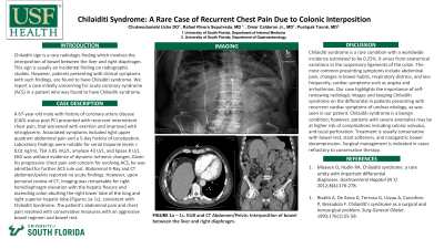
Case Description/Methods: A 67-year-old male with history of coronary artery disease (CAD) status post PCI presented with recurrent intermittent chest pain, that worsened with exertion and improved with nitroglycerin. Associated symptoms included right upper quadrant abdominal pain and a 5-day history of constipation. Laboratory findings were notable for serial troponin levels < 0.01 ng/ml, TSH 3.05 mU/L, amylase 43 U/L, and lipase 8 U/L. EKG was without evidence of dynamic ischemic changes. Given his progressive chest pain and concern for evolving ACS, he was admitted for further ACS rule out. Abdominal X-Ray and CT abdomen/pelvis reported no acute findings. However, upon personal review of CT, imaging was remarkable for right hemidiaphragm elevation with the hepatic flexure and ascending colon abutting the right lower lobe of the lung and right superior hepatic lobe (Figures 1a-1c), consistent with Chilaiditi Syndrome. The patient’s abdominal pain and chest pain resolved with conservative measures with an aggressive bowel regimen and bowel rest.
Discussion: Chilaiditi syndrome is a rare condition with a worldwide incidence estimated to be 0.25%. It arises from anatomical variations in the suspensory ligaments of the colon. The most common presenting symptoms include abdominal pain, changes in bowel habits, respiratory distress, and less frequently, cardiac symptoms such as angina and arrhythmias. Our case highlights the importance of self-reviewing radiologic images and keeping Chilaiditi syndrome on the differential in patients presenting with recurrent cardiac symptoms of unclear etiology, as was seen in our patient. Chilaiditi syndrome is a benign condition; however, patients with severe anomalies may be at higher risk of complications including colonic volvulus, and cecal perforation. Treatment is usually conservative with bowel rest, stool softeners, and nasogastric bowel decompression. Surgical management is indicated in cases refractory to conservative therapy.

Figure: 1a – 1c. KUB and CT Abdomen/Pelvis: Interposition of bowel between the liver and right diaphragm.
Disclosures:
Chukwudumebi Uche indicated no relevant financial relationships.
Rafael Rivera Sepulveda indicated no relevant financial relationships.
Omar Calderon indicated no relevant financial relationships.
Pushpak Taunk indicated no relevant financial relationships.
Chukwudumebi Uche, DO1, Rafael Rivera Sepulveda, MD1, Omar Calderon, MD1, Pushpak Taunk, MD2. P0289 - Chilaiditi Syndrome: A Rare Case of Recurrent Chest Pain Due to Colonic Interposition, ACG 2023 Annual Scientific Meeting Abstracts. Vancouver, BC, Canada: American College of Gastroenterology.


