Sunday Poster Session
Category: Colon
P0291 - A Rare Case of Amelanotic Rectal Melanoma
Sunday, October 22, 2023
3:30 PM - 7:00 PM PT
Location: Exhibit Hall

Has Audio

Sohil Khurana, DO
Northwell Health
Port Jefferson, NY
Presenting Author(s)
Sohil Khurana, DO1, Muhammad Jahanzaib Khan, MD2, Karina Fatakhova, MD2, Eugene Coman, MD1
1Northwell Health, Port Jefferson, NY; 2Mather Hospital/Northwell Health, Port Jefferson, NY
Introduction: Anorectal malignant melanoma (ARMM) is a rare aggressive tumor, with a dismal prognosis due to early metastasis. ARMM account for around 1% of all melanomas and anorectal tumors, with an average survival of 10 to 19 months after diagnosis. ARMM can be amelanotic in up to 30% of cases, and is often misdiagnosed as rectal adenocarcinoma, haemorrhoids, or benign polypoid lesions. We hereby present a rare case of amelanotic rectal melanoma.
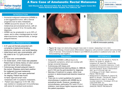
Case Description/Methods: A 61-year-old female with history of hypertension and invasive left sided lobular breast cancer status post bilateral mastectomy presented with complaints of abdominal bloating, obstructive defecation, and rectal bleeding. Initial blood work was unremarkable, with Hb noted to be 13.2 g/dl (normal 13-17 g/dL). A CT scan was performed showing diverticulosis, without any other acute findings. On rectal exam, a firm mass was palpated. The patient had no family history of colon cancer and had never had a colonoscopy. Subsequently, the patient underwent a colonoscopy, with a total of five polyps of varying sizes removed by polypectomy. Additionally, a large non-obstructing polypoid mass was seen in the rectum, measuring approximately 4 cm x 2cm, which was biopsied. Biopsies came back positive for malignant mucosal melanoma. An MRI was performed for staging, which demonstrated that the mass was 3.2 cm from anal verge and measured 2.5x2.5x1.9cm. The tumor was found to invade 6 mm into the upper right internal anal sphincter. No extra rectal extension was seen, but a few small suspicious mesorectal lymph nodes were noted. PET scan was performed showing FDG avidity in primary tumor site, and faint uptake in perirectal lymph nodes concerning for regional lymph node metastasis with no systemic metastasis. Brain MRI was also negative. Patient was staged as T2 N1 M0. She was started on neoadjuvant immunotherapy with plans for eventual local excision.
Discussion: Diagnosis of ARMM is difficult due to its nonspecific symptoms, which can overlap with benign anorectal diseases. Colonoscopy with biopsy and immunohistochemical staining (Melan-A, HMB-45, and S-100) are the gold standard for diagnosis. Mainstay of treatment is surgery, either local excision or abdominoperineal resection based on staging. There are no current guidelines for systemic chemotherapy in patients with ARMM due to suboptimal results. However, neoadjuvant immunotherapy, particularly PD-1 and CTLA-4 inhibitors, have shown higher response rates in this patient population.

Disclosures:
Sohil Khurana, DO1, Muhammad Jahanzaib Khan, MD2, Karina Fatakhova, MD2, Eugene Coman, MD1. P0291 - A Rare Case of Amelanotic Rectal Melanoma, ACG 2023 Annual Scientific Meeting Abstracts. Vancouver, BC, Canada: American College of Gastroenterology.
1Northwell Health, Port Jefferson, NY; 2Mather Hospital/Northwell Health, Port Jefferson, NY
Introduction: Anorectal malignant melanoma (ARMM) is a rare aggressive tumor, with a dismal prognosis due to early metastasis. ARMM account for around 1% of all melanomas and anorectal tumors, with an average survival of 10 to 19 months after diagnosis. ARMM can be amelanotic in up to 30% of cases, and is often misdiagnosed as rectal adenocarcinoma, haemorrhoids, or benign polypoid lesions. We hereby present a rare case of amelanotic rectal melanoma.
Case Description/Methods: A 61-year-old female with history of hypertension and invasive left sided lobular breast cancer status post bilateral mastectomy presented with complaints of abdominal bloating, obstructive defecation, and rectal bleeding. Initial blood work was unremarkable, with Hb noted to be 13.2 g/dl (normal 13-17 g/dL). A CT scan was performed showing diverticulosis, without any other acute findings. On rectal exam, a firm mass was palpated. The patient had no family history of colon cancer and had never had a colonoscopy. Subsequently, the patient underwent a colonoscopy, with a total of five polyps of varying sizes removed by polypectomy. Additionally, a large non-obstructing polypoid mass was seen in the rectum, measuring approximately 4 cm x 2cm, which was biopsied. Biopsies came back positive for malignant mucosal melanoma. An MRI was performed for staging, which demonstrated that the mass was 3.2 cm from anal verge and measured 2.5x2.5x1.9cm. The tumor was found to invade 6 mm into the upper right internal anal sphincter. No extra rectal extension was seen, but a few small suspicious mesorectal lymph nodes were noted. PET scan was performed showing FDG avidity in primary tumor site, and faint uptake in perirectal lymph nodes concerning for regional lymph node metastasis with no systemic metastasis. Brain MRI was also negative. Patient was staged as T2 N1 M0. She was started on neoadjuvant immunotherapy with plans for eventual local excision.
Discussion: Diagnosis of ARMM is difficult due to its nonspecific symptoms, which can overlap with benign anorectal diseases. Colonoscopy with biopsy and immunohistochemical staining (Melan-A, HMB-45, and S-100) are the gold standard for diagnosis. Mainstay of treatment is surgery, either local excision or abdominoperineal resection based on staging. There are no current guidelines for systemic chemotherapy in patients with ARMM due to suboptimal results. However, neoadjuvant immunotherapy, particularly PD-1 and CTLA-4 inhibitors, have shown higher response rates in this patient population.

Figure: A. Large non-obstructing polypoid mass was seen in the rectum, measuring roughly 4 cm x 2cm.
B. Histology showed poorly differentiated malignancy with prominent nucleoli and surrounding areas of necrosis. Immunostains for Melan-A and HMB-45 were positive in scattered tumor cells. S-100 immunostaining was negative. This is best classified as malignant melanoma.
B. Histology showed poorly differentiated malignancy with prominent nucleoli and surrounding areas of necrosis. Immunostains for Melan-A and HMB-45 were positive in scattered tumor cells. S-100 immunostaining was negative. This is best classified as malignant melanoma.
Disclosures:
Sohil Khurana indicated no relevant financial relationships.
Muhammad Jahanzaib Khan indicated no relevant financial relationships.
Karina Fatakhova indicated no relevant financial relationships.
Eugene Coman indicated no relevant financial relationships.
Sohil Khurana, DO1, Muhammad Jahanzaib Khan, MD2, Karina Fatakhova, MD2, Eugene Coman, MD1. P0291 - A Rare Case of Amelanotic Rectal Melanoma, ACG 2023 Annual Scientific Meeting Abstracts. Vancouver, BC, Canada: American College of Gastroenterology.
