Sunday Poster Session
Category: Colon
P0300 - A Rare Case of Retrorectal Cystic Hamartoma or Tailgut Cyst
Sunday, October 22, 2023
3:30 PM - 7:00 PM PT
Location: Exhibit Hall

Has Audio

Annie Shergill, MD
Larkin Community Hospital
Miami, FL
Presenting Author(s)
Annie Shergill, MD1, Annie Topham, MD2, Mahvish Khalid, MD3, Luis Geada, MD4, Luis Nasiff, MD2
1Larkin Community Hospital, Miami, FL; 2Larkin Community Hospital, Palm Springs Campus, Miami, FL; 3Larkin Community Hospital, Plam Springs Campus, Miami, FL; 4Baptist Health Hospital, Miami, FL
Introduction: A retrorectal cystic hamartoma or tailgut cyst (TGC) is an extremely rare congenital cystic lesion found in the presacral region. It is usually asymptomatic but patients may present with symptoms related to mass effect such as rectal fullness, constipation & chronic lower abdominal pain. We present a unique case of TGC diagnosed in a patient with acute diarrhea & infectious colitis.
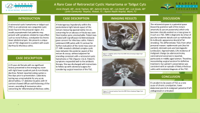
Case Description/Methods: A 25-year old female with no significant history presented to the emergency room with right lower quadrant pain & non-bloody diarrhea. Patient reported eating oysters a few days prior to presentation. Laboratory testing was unremarkable for any acute abnormalities. CT abdomen & pelvis with IV contrast showed diffuse wall thickening of the cecum, ascending & transverse colon concerning for inflammatory/infectious colitis. A heterogenous hypodensity within the posterolateral right lateral aspect of the rectum measuring approximately 3.6 cm, concerning for an abscess or fistula was seen. Stool studies were unremarkable. Patient was treated with Ciprofloxacin & Metronidazole given concern for infectious colitis. Patient underwent MRI pelvis with IV contrast for further evaluation of the rectal mass seen on CT. MRI revealed a bilobed complex cystic mass between the posterior aspect of the rectum & coccyx, without adjacent fat stranding consistent with a retrorectal cystic hamartoma or TGC. Patient’s symptoms responded well to the antibiotic therapy. She was discharged with instructions to follow-up with colorectal surgery for considering surgical resection of the TGC.
Discussion: The retrorectal space is a potential space (bound by posterior wall of the rectum anteriorly & sacrum posteriorly) which only becomes clinical evident as a mass grows in it such as a TGC. MRI is diagnostic by virtue of peculiar anatomic details such as multilocular & multicystic appearance devoid of fat stranding. This differentiates TGCs from other presacral masses- epidermoid cysts (has fat content), dermoid cysts and meningocele (unilocular). Approximately 13% incidence of malignant change (as adenocarcinoma, carcinoid and sarcomas) is reported, particularly in middle-aged women. Therefore, necessitating surgical excision for definitive treatment. Our patient’s presentation was consistent with an episode of likely infectious colitis with incidental diagnosis of the TGC. It's prudent to be aware of TGC as a rare cause of vague symptomatology such as chronic constipation, lower abdominal pain & its malignant potential.

Disclosures:
Annie Shergill, MD1, Annie Topham, MD2, Mahvish Khalid, MD3, Luis Geada, MD4, Luis Nasiff, MD2. P0300 - A Rare Case of Retrorectal Cystic Hamartoma or Tailgut Cyst, ACG 2023 Annual Scientific Meeting Abstracts. Vancouver, BC, Canada: American College of Gastroenterology.
1Larkin Community Hospital, Miami, FL; 2Larkin Community Hospital, Palm Springs Campus, Miami, FL; 3Larkin Community Hospital, Plam Springs Campus, Miami, FL; 4Baptist Health Hospital, Miami, FL
Introduction: A retrorectal cystic hamartoma or tailgut cyst (TGC) is an extremely rare congenital cystic lesion found in the presacral region. It is usually asymptomatic but patients may present with symptoms related to mass effect such as rectal fullness, constipation & chronic lower abdominal pain. We present a unique case of TGC diagnosed in a patient with acute diarrhea & infectious colitis.
Case Description/Methods: A 25-year old female with no significant history presented to the emergency room with right lower quadrant pain & non-bloody diarrhea. Patient reported eating oysters a few days prior to presentation. Laboratory testing was unremarkable for any acute abnormalities. CT abdomen & pelvis with IV contrast showed diffuse wall thickening of the cecum, ascending & transverse colon concerning for inflammatory/infectious colitis. A heterogenous hypodensity within the posterolateral right lateral aspect of the rectum measuring approximately 3.6 cm, concerning for an abscess or fistula was seen. Stool studies were unremarkable. Patient was treated with Ciprofloxacin & Metronidazole given concern for infectious colitis. Patient underwent MRI pelvis with IV contrast for further evaluation of the rectal mass seen on CT. MRI revealed a bilobed complex cystic mass between the posterior aspect of the rectum & coccyx, without adjacent fat stranding consistent with a retrorectal cystic hamartoma or TGC. Patient’s symptoms responded well to the antibiotic therapy. She was discharged with instructions to follow-up with colorectal surgery for considering surgical resection of the TGC.
Discussion: The retrorectal space is a potential space (bound by posterior wall of the rectum anteriorly & sacrum posteriorly) which only becomes clinical evident as a mass grows in it such as a TGC. MRI is diagnostic by virtue of peculiar anatomic details such as multilocular & multicystic appearance devoid of fat stranding. This differentiates TGCs from other presacral masses- epidermoid cysts (has fat content), dermoid cysts and meningocele (unilocular). Approximately 13% incidence of malignant change (as adenocarcinoma, carcinoid and sarcomas) is reported, particularly in middle-aged women. Therefore, necessitating surgical excision for definitive treatment. Our patient’s presentation was consistent with an episode of likely infectious colitis with incidental diagnosis of the TGC. It's prudent to be aware of TGC as a rare cause of vague symptomatology such as chronic constipation, lower abdominal pain & its malignant potential.

Figure: Sagittal section from the MRI pelvis showing the Tailgut Cyst (yellow arrow).
Disclosures:
Annie Shergill indicated no relevant financial relationships.
Annie Topham indicated no relevant financial relationships.
Mahvish Khalid indicated no relevant financial relationships.
Luis Geada indicated no relevant financial relationships.
Luis Nasiff indicated no relevant financial relationships.
Annie Shergill, MD1, Annie Topham, MD2, Mahvish Khalid, MD3, Luis Geada, MD4, Luis Nasiff, MD2. P0300 - A Rare Case of Retrorectal Cystic Hamartoma or Tailgut Cyst, ACG 2023 Annual Scientific Meeting Abstracts. Vancouver, BC, Canada: American College of Gastroenterology.
