Sunday Poster Session
Category: Colon
P0304 - A Rare Case of Appendiceal Inversion: Unraveling an Uncommon Condition
Sunday, October 22, 2023
3:30 PM - 7:00 PM PT
Location: Exhibit Hall

Has Audio
.jpg)
Rebhi Rabah, MD
SUNY Downstate Health Sciences University
Brooklyn, NY
Presenting Author(s)
Rebhi Rabah, MD, Aboud Kaliounji, MD, Justin Chung, MD, Narinderjeet Kaur, MS, MD, Michael Coles, MD, Evan Grossman, MD
SUNY Downstate Health Sciences University, Brooklyn, NY
Introduction: Appendiceal inversion (AI) is defined as an appendix that is pulled into the lumen of the cecum. It is a rare and harmless entity, incidentally found on a colonoscopy that presents a diagnostic dilemma to endoscopists as it can grossly resemble polypoid tissue.
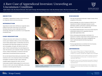
Case Description/Methods: A 69-year-old woman with a past medical history of iron deficiency anemia, type II diabetes mellitus, coronary artery disease, chronic kidney disease stage V, and blindness was sent to the hospital after labs revealed her to be anemic, with a hemoglobin of 5.2 g/dL, MCV 65.9 fL, ferritin of 11 ng/mL. Upon initial assessment, patient endorsed feeling chronically fatigued and weak. She denied any prior episodes of bright red blood per rectum, melena, hematemesis, or hematuria. She had a positive fecal immunochemical test a year prior presentation, which was not followed up with a colonoscopy. She was hemodynamically stable upon presentation. After transfusion with 2 units of packed red blood cells (pRBC), her hemoglobin responded appropriately and increased to 8.1 g/dL. An upper endoscopy was performed and revealed 3 gastric polyps, 2 of them being 15 mm and one 40 mm, while a colonoscopy revealed 2 sessile polyps, 2 mm and 5 mm in size, in the cecum all of which were removed. Additionally, her colonoscopy also revealed an inverted appendix (Figure 1).
Discussion: While exceedingly rare with an incidence of 0.01%, it is imperative to be able distinguish between AI and a pathologic condition- particularly in patients with risk factors for colon neoplasms or previous polyps. Any polypoid appearing lesion in the cecum should raise the endoscopist's suspicion of AI, especially in the absence of an appreciable appendiceal orifice. Neglecting to maintain a high index of clinical suspicion for AI during endoscopy with cecal lesions may lead to unnecessary and invasive investigations resulting in perforation/peritonitis following biopsy or transection. This abstract and associated endoscopic images serves to increase clinical recognition of this entity. Additionally, previous appendectomy is considered a risk factor for AI for which our patient never had, highlighting no definitive patient phenotype exists for the presence of AI.

Disclosures:
Rebhi Rabah, MD, Aboud Kaliounji, MD, Justin Chung, MD, Narinderjeet Kaur, MS, MD, Michael Coles, MD, Evan Grossman, MD. P0304 - A Rare Case of Appendiceal Inversion: Unraveling an Uncommon Condition, ACG 2023 Annual Scientific Meeting Abstracts. Vancouver, BC, Canada: American College of Gastroenterology.
SUNY Downstate Health Sciences University, Brooklyn, NY
Introduction: Appendiceal inversion (AI) is defined as an appendix that is pulled into the lumen of the cecum. It is a rare and harmless entity, incidentally found on a colonoscopy that presents a diagnostic dilemma to endoscopists as it can grossly resemble polypoid tissue.
Case Description/Methods: A 69-year-old woman with a past medical history of iron deficiency anemia, type II diabetes mellitus, coronary artery disease, chronic kidney disease stage V, and blindness was sent to the hospital after labs revealed her to be anemic, with a hemoglobin of 5.2 g/dL, MCV 65.9 fL, ferritin of 11 ng/mL. Upon initial assessment, patient endorsed feeling chronically fatigued and weak. She denied any prior episodes of bright red blood per rectum, melena, hematemesis, or hematuria. She had a positive fecal immunochemical test a year prior presentation, which was not followed up with a colonoscopy. She was hemodynamically stable upon presentation. After transfusion with 2 units of packed red blood cells (pRBC), her hemoglobin responded appropriately and increased to 8.1 g/dL. An upper endoscopy was performed and revealed 3 gastric polyps, 2 of them being 15 mm and one 40 mm, while a colonoscopy revealed 2 sessile polyps, 2 mm and 5 mm in size, in the cecum all of which were removed. Additionally, her colonoscopy also revealed an inverted appendix (Figure 1).
Discussion: While exceedingly rare with an incidence of 0.01%, it is imperative to be able distinguish between AI and a pathologic condition- particularly in patients with risk factors for colon neoplasms or previous polyps. Any polypoid appearing lesion in the cecum should raise the endoscopist's suspicion of AI, especially in the absence of an appreciable appendiceal orifice. Neglecting to maintain a high index of clinical suspicion for AI during endoscopy with cecal lesions may lead to unnecessary and invasive investigations resulting in perforation/peritonitis following biopsy or transection. This abstract and associated endoscopic images serves to increase clinical recognition of this entity. Additionally, previous appendectomy is considered a risk factor for AI for which our patient never had, highlighting no definitive patient phenotype exists for the presence of AI.

Figure: Figure 1: Image A and B showing inverted appendix resembling polypoid tissue.
Disclosures:
Rebhi Rabah indicated no relevant financial relationships.
Aboud Kaliounji indicated no relevant financial relationships.
Justin Chung indicated no relevant financial relationships.
Narinderjeet Kaur indicated no relevant financial relationships.
Michael Coles indicated no relevant financial relationships.
Evan Grossman indicated no relevant financial relationships.
Rebhi Rabah, MD, Aboud Kaliounji, MD, Justin Chung, MD, Narinderjeet Kaur, MS, MD, Michael Coles, MD, Evan Grossman, MD. P0304 - A Rare Case of Appendiceal Inversion: Unraveling an Uncommon Condition, ACG 2023 Annual Scientific Meeting Abstracts. Vancouver, BC, Canada: American College of Gastroenterology.
