Sunday Poster Session
Category: Endoscopy Video Forum
P0377 - Periampullary Diverticulum Obscuring Visualization of a Type III Choledochal Cyst: A Rare Presentation

.jpg)
Albert Manudhane, MD
The Ohio State University Wexner Medical Center
Columbus, OH
Presenting Author(s)
1The Ohio State University Wexner Medical Center, Columbus, OH; 2The Ohio State Wexner Medical Center, Columbus, OH
Introduction:
Choledochal cysts (CDCs) are infrequently occurring inborn dilations of the extrahepatic or intrahepatic ducts, with a frequency of 1 in 100,000 births among Westerners. This abnormality is more frequently seen in the Asian population and in females, with a majority of cases diagnosed in childhood. Among the various types of CDCs classified by Todani, type III CDCs, also known as choledochoceles, are the least common, accounting for 1.4-4.5% of all cases. These cystic dilations occur in the distal common bile duct and can protrude into the duodenum. In this report, we present a unique case of a type III CDC adjacent to a periampullary diverticulum.
Case Description/Methods:
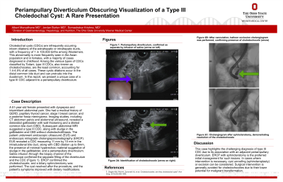
A 51-year-old female presented with dyspepsia and intermittent abdominal pain. She had a medical history of GERD, papillary thyroid cancer, stage I breast cancer, and a posterior fossa meningioma. Imaging studies, including CT abdomen pelvis and abdominal ultrasound, revealed a distended gallbladder with wall thickening and a dilated common bile duct (CBD). Subsequent abdominal MRI suggested a type III CDC, along with sludge in the gallbladder and CBD without choledocholithiasis. The patient underwent endoscopic ultrasound (EUS) and endoscopic retrograde cholangiopancreatography (ERCP). EUS revealed a CDC measuring 11 mm by 10 mm in the intraduodenal bile duct, along with CBD dilation up to 8mm, the presence of minimal hyperechoic material suggestive of sludge in the gallbladder, and a periampullary diverticulum. Saline infusion through the biopsy channel of the endoscope confirmed the separate filling of the diverticulum and the CDC (Video 1). ERCP confirmed the choledochocele, and a biliary sphincterotomy was performed. The cyst resolved after the procedure, and the patient's symptoms improved with dietary modifications.
Discussion:
This case highlights the challenging diagnosis of type III CDC due to its association with an adjacent periampullary diverticulum. ERCP with sphincterotomy is the preferred initial management for such lesions. In cases where intervention is necessary, cyst unroofing (sphincteroplasty) or excision can be considered. Surgical intervention is generally avoided for choledochoceles due to their lower potential for malignant transformation.
Disclosures:
Albert Manudhane, MD1, Jordan Burlen, MD1, Somashekar Krishna, MD2. P0377 - Periampullary Diverticulum Obscuring Visualization of a Type III Choledochal Cyst: A Rare Presentation, ACG 2023 Annual Scientific Meeting Abstracts. Vancouver, BC, Canada: American College of Gastroenterology.
