Sunday Poster Session
Category: Endoscopy Video Forum
P0378 - Hemosuccus Pancreaticus From the Minor Papilla: A Rare Cause of Upper GI Bleed
Sunday, October 22, 2023
3:30 PM - 7:00 PM PT
Location: Exhibit Hall

Has Audio
- NR
Nikhil Reddy, MD
University of Chicago
Chicago, IL
Presenting Author(s)
Nikhil Reddy, MD1, Grace E.. Kim, MD1, Mary Ryan, MD1, Sajan Nagpal, MD2, Uzma D. Siddiqui, MD3
1University of Chicago, Chicago, IL; 2MNGI Digestive Health, Eagan, MN; 3Center for Endoscopic Research and Therapeutics (CERT), University of Chicago, Chicago, IL
Introduction: Hemosuccus pancreaticus is a rare cause of upper gastrointestinal (GI) bleeding, accounting for less than one in 1,500 cases. It most frequently occurs through the major papilla, but less commonly, bleeding can occur out of the minor papilla, a condition only described in a few case reports. Here, we describe a case of a 59-year-old man with a history of alcoholic cirrhosis, esophageal varices, and pancreatic lesions who experienced hematemesis and bleeding from the minor papilla which was visualized on side-viewing duodenoscope.
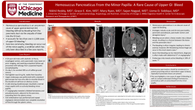
Case Description/Methods: This case involves a 59-year-old man with a history of alcoholic cirrhosis, esophageal varices, and a pancreatic mass (seen on prior imaging, and awaiting outpatient follow-up) who was admitted with lethargy from suspected hepatic encephalopathy. On day 3, he vomited 300 ccs of coffee-ground emesis. Physical exam was significant for epigastric tenderness. Laboratory results showed a hemoglobin of 8.4 g/dL, stable from baseline. Upper endoscopy was performed with a standard gastroscope but was only able to visualize blood in the duodenum without an obvious source. Next, a side viewing duodenoscope visualized a normal major papilla. Subsequently, active bleeding from the minor papilla was identified. CT Angiography revealed a bilobed hematoma and pseudoaneurysm, adjacent to the pancreas, extending from the splenic artery. Coil embolization was performed and repeat angiogram demonstrated near complete resolution of flow through the splenic artery.
Discussion: Hemosuccus pancreaticus is an obscure cause of upper GI bleeding defined by bleeding from the pancreatic duct via the ampulla of Vater into the GI tract. A rare subgroup of this already uncommon condition occurs when bleeding transpires through the minor papilla. Bleeding occurs when a lesion erodes into a blood vessel, resulting in a channel between the lesion and the pancreatic duct. The bleeding is often intermittent, leading to chronic anemia; however, life-threatening hemorrhage can occur in the most severe cases. Given that bleeding can be irregular, diagnosis is often made with abdominal CT or angiography. Endoscopy only reveals active bleeding in 30% of cases. Overall, this case illustrates an extremely rare cause of upper GI bleeding: hemosuccus pancreaticus with active bleeding from the minor papilla, visualized on endoscopy.

Disclosures:
Nikhil Reddy, MD1, Grace E.. Kim, MD1, Mary Ryan, MD1, Sajan Nagpal, MD2, Uzma D. Siddiqui, MD3. P0378 - Hemosuccus Pancreaticus From the Minor Papilla: A Rare Cause of Upper GI Bleed, ACG 2023 Annual Scientific Meeting Abstracts. Vancouver, BC, Canada: American College of Gastroenterology.
1University of Chicago, Chicago, IL; 2MNGI Digestive Health, Eagan, MN; 3Center for Endoscopic Research and Therapeutics (CERT), University of Chicago, Chicago, IL
Introduction: Hemosuccus pancreaticus is a rare cause of upper gastrointestinal (GI) bleeding, accounting for less than one in 1,500 cases. It most frequently occurs through the major papilla, but less commonly, bleeding can occur out of the minor papilla, a condition only described in a few case reports. Here, we describe a case of a 59-year-old man with a history of alcoholic cirrhosis, esophageal varices, and pancreatic lesions who experienced hematemesis and bleeding from the minor papilla which was visualized on side-viewing duodenoscope.
Case Description/Methods: This case involves a 59-year-old man with a history of alcoholic cirrhosis, esophageal varices, and a pancreatic mass (seen on prior imaging, and awaiting outpatient follow-up) who was admitted with lethargy from suspected hepatic encephalopathy. On day 3, he vomited 300 ccs of coffee-ground emesis. Physical exam was significant for epigastric tenderness. Laboratory results showed a hemoglobin of 8.4 g/dL, stable from baseline. Upper endoscopy was performed with a standard gastroscope but was only able to visualize blood in the duodenum without an obvious source. Next, a side viewing duodenoscope visualized a normal major papilla. Subsequently, active bleeding from the minor papilla was identified. CT Angiography revealed a bilobed hematoma and pseudoaneurysm, adjacent to the pancreas, extending from the splenic artery. Coil embolization was performed and repeat angiogram demonstrated near complete resolution of flow through the splenic artery.
Discussion: Hemosuccus pancreaticus is an obscure cause of upper GI bleeding defined by bleeding from the pancreatic duct via the ampulla of Vater into the GI tract. A rare subgroup of this already uncommon condition occurs when bleeding transpires through the minor papilla. Bleeding occurs when a lesion erodes into a blood vessel, resulting in a channel between the lesion and the pancreatic duct. The bleeding is often intermittent, leading to chronic anemia; however, life-threatening hemorrhage can occur in the most severe cases. Given that bleeding can be irregular, diagnosis is often made with abdominal CT or angiography. Endoscopy only reveals active bleeding in 30% of cases. Overall, this case illustrates an extremely rare cause of upper GI bleeding: hemosuccus pancreaticus with active bleeding from the minor papilla, visualized on endoscopy.

Figure: A: CTA demonstrating a pseudoaneurysm originating from the splenic artery measuring 2.4 x 1.6 cm (red arrow)
B: CTA showing a bilobed hematoma associated with the pancreas measuring 5.8 x 5.4 cm at the head of the pancreas (red circle)
C: Pre-embolization angiogram revealing a patent splenic artery (red arrow)
D: Post-embolization angiogram demonstrating resolution of flow through the proximal splenic artery (red arrow)
Disclosures:
Nikhil Reddy indicated no relevant financial relationships.
Grace Kim indicated no relevant financial relationships.
Mary Ryan indicated no relevant financial relationships.
Sajan Nagpal: Nestle Health Sciences – Speakers Bureau.
Uzma Siddiqui: Boston Scientific – Consultant, Grant/Research Support, Speaker. ConMed – Consultant, Speaker. Cook – Consultant, Speaker. Medtronic – Consultant, Speaker. Olympus – Consultant, Grant/Research Support, Speaker.
Nikhil Reddy, MD1, Grace E.. Kim, MD1, Mary Ryan, MD1, Sajan Nagpal, MD2, Uzma D. Siddiqui, MD3. P0378 - Hemosuccus Pancreaticus From the Minor Papilla: A Rare Cause of Upper GI Bleed, ACG 2023 Annual Scientific Meeting Abstracts. Vancouver, BC, Canada: American College of Gastroenterology.

