Sunday Poster Session
Category: Endoscopy Video Forum
P0380 - Management of Choledocholithiasis in a Complex Surgical Abdomen Pushed to the EDGE


Kristelle Imperio-Lagabon, MD
Cleveland Clinic Foundation
Cleveland Heights, OH
Presenting Author(s)
1Cleveland Clinic Foundation, Cleveland Heights, OH; 2Cleveland Clinic Foundation, Cleveland, OH; 3Cleveland Clinic, Cleveland, OH
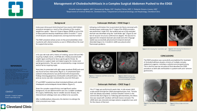
Introduction: Endoscopic Ultrasound Directed Trans-Gastric ERCP (EDGE) procedure emerged due to the rising number of people undergoing a Roux-en-Y Gastric Bypass (RYGB). The EDGE procedure allows access to the pancreaticobiliary trees in those with altered anatomy as seen in RYGB without need for surgical intervention.
Case Description/Methods: A 61-year-old male with RYGB had weight regain and was found to have a gastro-gastric fistula. Revision with remnant subtotal gastrectomy complicated by obstruction requiring a repeat revision and later a hernia repair. He later presented with right upper quadrant abdominal (RUQ) pain, found to have cholecystitis. A complicated open subtotal cholecystectomy was performed showing gangrenous cholecystitis with perforation. The postoperative course was complicated by persistent RUQ abdominal pain. Imaging revealed new distal choledocholithiasis on CT and MRI. Given complex surgical history with significant cardiac disease, he was deemed a poor surgical candidate. His notable vascular disease made interventional radiology unlikely to be successful. Thus the decision to undergo EDGE was made.
In the first stage of the EDGE procedure, the remnant stomach was identified, under ultrasound guidance, by the "sand dollar sign". Once the lack of vascular structures were confirmed via doppler, a 19-gauge SharkCore needle was advanced through the jejunum to the remnant stomach and distended with 500cc of a mixed solution. A jejunogastrostomy was made by electrocautery device to increase stoma size and a 15x10 mm AXIOS stent was placed.
The second stage was performed 1 month later. A side-viewer ERCP scope passed through the AXIOS jejunogastrostomy tract. The bile duct was deeply cannulated with the short-nosed traction sphincterotome and guidewire. Contrast was injected, revealing two stones in the common bile duct. Following sphincterotomy, the biliary tree was swept with an 8.5mm and 11.5mm balloon with sludge swept and both stones removed. The AXIOS stent was removed using a Raptor grasping device and fulguration using argon plasma at 0.8 L/min and 20 watts used for closure of the tract.
Discussion: The EDGE procedure was successfully accomplished for treatment of choledocholithiasis despite a history of multiple complex abdominal surgeries including subtotal remnant gastrectomy. This is the first case we are aware of describing the EDGE procedure as both safe and feasible in a patient with a prior remnant gastrectomy.
Disclosures:
Kristelle Imperio-Lagabon, MD1, Stephen A. Firkins, MD2, Ramanpreet Bajwa, DO2, C. Roberto Simons-Linares, MD3. P0380 - Management of Choledocholithiasis in a Complex Surgical Abdomen Pushed to the EDGE, ACG 2023 Annual Scientific Meeting Abstracts. Vancouver, BC, Canada: American College of Gastroenterology.
