Sunday Poster Session
Category: Endoscopy Video Forum
P0381 - EUS-Guided Confocal Laser Endomicroscopy-Based Differentiation of Dysplasia in Intraductal Papillary Mucinous Neoplasms
Sunday, October 22, 2023
3:30 PM - 7:00 PM PT
Location: Exhibit Hall

Has Audio

Da Yeon Ryoo, MD
Ohio State University Wexner Medical Center
Columbus, OH
Presenting Author(s)
Da Yeon Ryoo, MD1, Bryn Koehler, MD1, Matt Leupold, MD2, Troy Cao, BS1, Wei Chen, MD, PhD1, Somashekar Krishna, MD3
1Ohio State University Wexner Medical Center, Columbus, OH; 2The Ohio State University Wexner Medical Center, Columbus, OH; 3The Ohio State Wexner Medical Center, Columbus, OH
Introduction: Endoscopic ultrasound (EUS) guided needle-based confocal laser endomicroscopy (nCLE) offers real-time, in vivo microscopic visualization of intraductal papillary mucinous neoplasms (IPMNs). In this video, we provide a detailed description of the features that distinguish between branch duct (BD) IPMNs with low-grade dysplasia (LGD) and those with high-grade dysplasia and adenocarcinoma (HGD-Ca).
Case Description/Methods: The EUS was conducted using a linear echoendoscope. A 19-gauge needle, pre-loaded with the AQ-Flex nCLE mini-probe (Cellvizio, Mauna Kea Technologies, Paris, France), was inserted into the pancreatic cystic lesion (PCL) under EUS guidance. The mini-probe was advanced beyond the needle tip to obtain endomicroscopy imaging. The endomicroscopy imaging was performed with permissive angulation of the needle, elevator, and rotation around the operator's body axis. Selected patients in this series subsequently underwent surgical resection of the PCL, and their histopathological report was utilized as the reference standard.
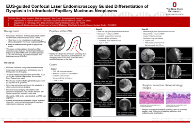
Discussion: The video features three selected cases: one low-grade dysplasia (LGD) IPMN and two IPMNs with high-grade dysplasia/adenocarcinoma (HGD-Ca). In the videos, the papillae within the PCL are easily noticeable. These papillae are finger-like structures consisting of an inner vascular core and an outer epithelium.
Progressive dysplastic changes result in an increase in the size of the papillae and a thickening of the papillary epithelium, caused by cellular and nuclear stratification. The stratification of nuclei imparts a darker hue, leading to increased darkness of the epithelium. The papillae in the IPMN with LGD exhibit a thinner and more translucent epithelium, whereas the IPMN with HGD-Ca displays thicker and darker papillae that are also larger in size.
Other notable changes associated with progressive dysplasia include the loss of papillary architecture with irregular borders and the disappearance of the vascular core (or lamina propria). In some cases, as demonstrated in the video featuring HGD-Ca, large cells (~30-50 microns) with prominent nuclei indicative of adenocarcinoma can be observed. The corresponding histopathology images from two of the selected cases serve as the reference standard, confirming these observed changes.
In summary, EUS-nCLE provides the ability to differentiate and characterize IPMNs through endomicroscopic visualization. This method holds great promise as a potential modality for the management of BD-IPMNs.
Disclosures:
Da Yeon Ryoo, MD1, Bryn Koehler, MD1, Matt Leupold, MD2, Troy Cao, BS1, Wei Chen, MD, PhD1, Somashekar Krishna, MD3. P0381 - EUS-Guided Confocal Laser Endomicroscopy-Based Differentiation of Dysplasia in Intraductal Papillary Mucinous Neoplasms, ACG 2023 Annual Scientific Meeting Abstracts. Vancouver, BC, Canada: American College of Gastroenterology.
1Ohio State University Wexner Medical Center, Columbus, OH; 2The Ohio State University Wexner Medical Center, Columbus, OH; 3The Ohio State Wexner Medical Center, Columbus, OH
Introduction: Endoscopic ultrasound (EUS) guided needle-based confocal laser endomicroscopy (nCLE) offers real-time, in vivo microscopic visualization of intraductal papillary mucinous neoplasms (IPMNs). In this video, we provide a detailed description of the features that distinguish between branch duct (BD) IPMNs with low-grade dysplasia (LGD) and those with high-grade dysplasia and adenocarcinoma (HGD-Ca).
Case Description/Methods: The EUS was conducted using a linear echoendoscope. A 19-gauge needle, pre-loaded with the AQ-Flex nCLE mini-probe (Cellvizio, Mauna Kea Technologies, Paris, France), was inserted into the pancreatic cystic lesion (PCL) under EUS guidance. The mini-probe was advanced beyond the needle tip to obtain endomicroscopy imaging. The endomicroscopy imaging was performed with permissive angulation of the needle, elevator, and rotation around the operator's body axis. Selected patients in this series subsequently underwent surgical resection of the PCL, and their histopathological report was utilized as the reference standard.
Discussion: The video features three selected cases: one low-grade dysplasia (LGD) IPMN and two IPMNs with high-grade dysplasia/adenocarcinoma (HGD-Ca). In the videos, the papillae within the PCL are easily noticeable. These papillae are finger-like structures consisting of an inner vascular core and an outer epithelium.
Progressive dysplastic changes result in an increase in the size of the papillae and a thickening of the papillary epithelium, caused by cellular and nuclear stratification. The stratification of nuclei imparts a darker hue, leading to increased darkness of the epithelium. The papillae in the IPMN with LGD exhibit a thinner and more translucent epithelium, whereas the IPMN with HGD-Ca displays thicker and darker papillae that are also larger in size.
Other notable changes associated with progressive dysplasia include the loss of papillary architecture with irregular borders and the disappearance of the vascular core (or lamina propria). In some cases, as demonstrated in the video featuring HGD-Ca, large cells (~30-50 microns) with prominent nuclei indicative of adenocarcinoma can be observed. The corresponding histopathology images from two of the selected cases serve as the reference standard, confirming these observed changes.
In summary, EUS-nCLE provides the ability to differentiate and characterize IPMNs through endomicroscopic visualization. This method holds great promise as a potential modality for the management of BD-IPMNs.
Disclosures:
Da Yeon Ryoo indicated no relevant financial relationships.
Bryn Koehler indicated no relevant financial relationships.
Matt Leupold indicated no relevant financial relationships.
Troy Cao indicated no relevant financial relationships.
Wei Chen indicated no relevant financial relationships.
Somashekar Krishna: Mauna Kea Technologies – Grant/Research Support. Taewoong Medical USA – Grant/Research Support. US Biotest – Grant/Research Support.
Da Yeon Ryoo, MD1, Bryn Koehler, MD1, Matt Leupold, MD2, Troy Cao, BS1, Wei Chen, MD, PhD1, Somashekar Krishna, MD3. P0381 - EUS-Guided Confocal Laser Endomicroscopy-Based Differentiation of Dysplasia in Intraductal Papillary Mucinous Neoplasms, ACG 2023 Annual Scientific Meeting Abstracts. Vancouver, BC, Canada: American College of Gastroenterology.
