Sunday Poster Session
Category: Endoscopy Video Forum
P0383 - ERCP for Postoperative Bile Leak
Sunday, October 22, 2023
3:30 PM - 7:00 PM PT
Location: Exhibit Hall

Has Audio

Deepak K. Johnson, MD, DM
BCMCH
Thiruvalla, Kerala, India
Presenting Author(s)
Deepak K. Johnson, MD, DM
BCMCH, Thiruvalla, Kerala, India
Introduction: We hereby present a patient who developed a disastrous complication of Whipples pancreato duodenectomy.The situation was salvaged by a combination of interventional radiology + Endoscopic (ERCP) interventions
Case Description/Methods: Fifty one year old male presented with Upper abdominal pain + weight loss over 6 months.Liver function tests showed cholestatic pattern and CA 19 -9 was elevated (223 U/ml).CECT abdomen showed chronic pancreatitis with a 4 cm mass in head-uncinate with biliary obstruction and multiple liver hemangiomas.Patient underwent open Whipples pancreatoduodenectomy .
Patient developed severe abdominal pain on POD5.Imaging showed large subdiaphragmatic collection. Percutaneous drain was placed by radiologist and oral feeds started. However the drain slipped out on POD8- Patient was shifted to ICU in view of severe abdominal pain and tachycardia and drain was re inserted. Bile refeeding was done later daily along with oral diet intake. Interval imaging showed reduction in bile collection size. Endoscopic biliary stenting was planned on POD 21.
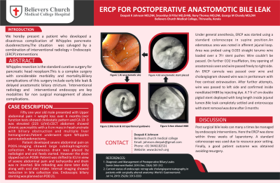
Under general anesthesia, ERCP was started using a standard colonoscope in supine position. Afferent loop was entered and confirmed by presence of bile and movement of scope towards right upper quadrant in fluoro. Subsequently, an edematous area was noted which could be the site of hepaticojejunostomy. Also scope tip was seen close to surgical clips in fluoro. Area was probed using 0.035 straight terumo wire loaded over a 7Fr stent pusher. Wire couldn’t be passed. On further CO2 insufflation, tiny opening of anastomosis seen and wire passed freely to right side. An ERCP cannula was passed over wire and cholangiogram showed wire was in peritoneum with free contrast extravasation. After further attempts, wire was passed to left side and confirmed inside nondilated IHBR by injecting dye. A 7 fr x7 cm double pigtail stent deployed with long length inside jejunal lumen.
During follow up, bile leak completely settled and enteroscopy with stent removal was done after 3 months. Patient is doing well during follow up.The biopsy showed that the pancreatic head lesion was an inflammatory one in nature with changes of underlying chronic pancreatitis.
Discussion: Post surgical bile leaks can many a times be managed by endoscopic interventions. Here the ERCP was done within three weeks of laparotomy. A standard colonoscope was used due to resource poor setting. Finally, a good patient outcome was obtained avoiding resurgery
Disclosures:
Deepak K. Johnson, MD, DM. P0383 - ERCP for Postoperative Bile Leak, ACG 2023 Annual Scientific Meeting Abstracts. Vancouver, BC, Canada: American College of Gastroenterology.
BCMCH, Thiruvalla, Kerala, India
Introduction: We hereby present a patient who developed a disastrous complication of Whipples pancreato duodenectomy.The situation was salvaged by a combination of interventional radiology + Endoscopic (ERCP) interventions
Case Description/Methods: Fifty one year old male presented with Upper abdominal pain + weight loss over 6 months.Liver function tests showed cholestatic pattern and CA 19 -9 was elevated (223 U/ml).CECT abdomen showed chronic pancreatitis with a 4 cm mass in head-uncinate with biliary obstruction and multiple liver hemangiomas.Patient underwent open Whipples pancreatoduodenectomy .
Patient developed severe abdominal pain on POD5.Imaging showed large subdiaphragmatic collection. Percutaneous drain was placed by radiologist and oral feeds started. However the drain slipped out on POD8- Patient was shifted to ICU in view of severe abdominal pain and tachycardia and drain was re inserted. Bile refeeding was done later daily along with oral diet intake. Interval imaging showed reduction in bile collection size. Endoscopic biliary stenting was planned on POD 21.
Under general anesthesia, ERCP was started using a standard colonoscope in supine position. Afferent loop was entered and confirmed by presence of bile and movement of scope towards right upper quadrant in fluoro. Subsequently, an edematous area was noted which could be the site of hepaticojejunostomy. Also scope tip was seen close to surgical clips in fluoro. Area was probed using 0.035 straight terumo wire loaded over a 7Fr stent pusher. Wire couldn’t be passed. On further CO2 insufflation, tiny opening of anastomosis seen and wire passed freely to right side. An ERCP cannula was passed over wire and cholangiogram showed wire was in peritoneum with free contrast extravasation. After further attempts, wire was passed to left side and confirmed inside nondilated IHBR by injecting dye. A 7 fr x7 cm double pigtail stent deployed with long length inside jejunal lumen.
During follow up, bile leak completely settled and enteroscopy with stent removal was done after 3 months. Patient is doing well during follow up.The biopsy showed that the pancreatic head lesion was an inflammatory one in nature with changes of underlying chronic pancreatitis.
Discussion: Post surgical bile leaks can many a times be managed by endoscopic interventions. Here the ERCP was done within three weeks of laparotomy. A standard colonoscope was used due to resource poor setting. Finally, a good patient outcome was obtained avoiding resurgery
Disclosures:
Deepak Johnson indicated no relevant financial relationships.
Deepak K. Johnson, MD, DM. P0383 - ERCP for Postoperative Bile Leak, ACG 2023 Annual Scientific Meeting Abstracts. Vancouver, BC, Canada: American College of Gastroenterology.

