Sunday Poster Session
Category: Endoscopy Video Forum
P0385 - Management of a Rare Complication of a Percutaneous Gastrostomy Tube Placement
Sunday, October 22, 2023
3:30 PM - 7:00 PM PT
Location: Exhibit Hall

Has Audio

Adrian Pona, MD
Prisma Health-Upstate
Greenville, SC
Presenting Author(s)
Adrian Pona, MD1, Varun Moktan, MD1, Einar Lurix, MD2, Lance Mcleroy, MD2, Thomas Dersch, MD2, Rupal Patel, MD2, Veeral M. Oza, MD2
1Prisma Health-Upstate, Greenville, SC; 2Prisma Health, Greenville, SC
Introduction: Endoscopic ultrasound (EUS) is a valuable tool in diagnosing and managing gastrointestinal disorders. However, its role in addressing uncommon scenarios such as post-surgical complications remain unanswered. We present a case highlighting the innovative use of EUS in managing a complication following percutaneous endoscopic gastrostomy (PEG) placement.
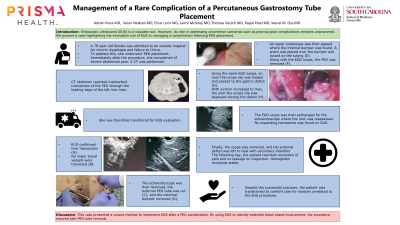
Case Description/Methods: A 78-year-old female was admitted to an outside hospital for chronic dysphagia and failure to thrive. To address her chronic dysphagia, and failure to thrive, she underwent a PEG placement. Immediately after the PEG placement she complained of severe abdominal pain. She continued to report significant pain one day post-procedure. A CT scan revealed inadvertent placement of the PEG through the leading edge of the left lobe of the liver. The patient was subsequently transferred for possible endoscopic management of this complication in lieu of a surgery. It was decided to perform an EUS to evaluate any potential blood vessel involvement by the misplaced PEG tube. Using a linear echoendoscope, the PEG was identified in the stomach, and the course of the PEG was also clearly noted to be through the left lobe of the liver. Major blood vessel involvement was not noted. The external PEG tube was then cut, and the external bumper was removed. An upper endoscope was then used to locate and snare the internal bumper. The tubing was gently pulled through the defect, and an over-the-scope clip was deployed to close the gastric defect. Both internal and external examination confirmed successful closure. The upper endoscope was exchanged for the linear echoendoscope where the liver was once again assessed. No expanding hematomas were noted on EUS exam. The next day, the patient reported near complete resolution of abdominal pain and no leakage was found on inspection. Her hemoglobin also remained stable.
Discussion: This case report presents a unique way to implement EUS after a PEG complication. By using EUS to identify potential blood vessels involvement, the procedure ensured safe PEG tube removal with a low bleeding risk. Endoscopic ultrasound indications and usage continues to grow and this case is an example of such a use.
Disclosures:
Adrian Pona, MD1, Varun Moktan, MD1, Einar Lurix, MD2, Lance Mcleroy, MD2, Thomas Dersch, MD2, Rupal Patel, MD2, Veeral M. Oza, MD2. P0385 - Management of a Rare Complication of a Percutaneous Gastrostomy Tube Placement, ACG 2023 Annual Scientific Meeting Abstracts. Vancouver, BC, Canada: American College of Gastroenterology.
1Prisma Health-Upstate, Greenville, SC; 2Prisma Health, Greenville, SC
Introduction: Endoscopic ultrasound (EUS) is a valuable tool in diagnosing and managing gastrointestinal disorders. However, its role in addressing uncommon scenarios such as post-surgical complications remain unanswered. We present a case highlighting the innovative use of EUS in managing a complication following percutaneous endoscopic gastrostomy (PEG) placement.
Case Description/Methods: A 78-year-old female was admitted to an outside hospital for chronic dysphagia and failure to thrive. To address her chronic dysphagia, and failure to thrive, she underwent a PEG placement. Immediately after the PEG placement she complained of severe abdominal pain. She continued to report significant pain one day post-procedure. A CT scan revealed inadvertent placement of the PEG through the leading edge of the left lobe of the liver. The patient was subsequently transferred for possible endoscopic management of this complication in lieu of a surgery. It was decided to perform an EUS to evaluate any potential blood vessel involvement by the misplaced PEG tube. Using a linear echoendoscope, the PEG was identified in the stomach, and the course of the PEG was also clearly noted to be through the left lobe of the liver. Major blood vessel involvement was not noted. The external PEG tube was then cut, and the external bumper was removed. An upper endoscope was then used to locate and snare the internal bumper. The tubing was gently pulled through the defect, and an over-the-scope clip was deployed to close the gastric defect. Both internal and external examination confirmed successful closure. The upper endoscope was exchanged for the linear echoendoscope where the liver was once again assessed. No expanding hematomas were noted on EUS exam. The next day, the patient reported near complete resolution of abdominal pain and no leakage was found on inspection. Her hemoglobin also remained stable.
Discussion: This case report presents a unique way to implement EUS after a PEG complication. By using EUS to identify potential blood vessels involvement, the procedure ensured safe PEG tube removal with a low bleeding risk. Endoscopic ultrasound indications and usage continues to grow and this case is an example of such a use.
Disclosures:
Adrian Pona indicated no relevant financial relationships.
Varun Moktan indicated no relevant financial relationships.
Einar Lurix indicated no relevant financial relationships.
Lance Mcleroy indicated no relevant financial relationships.
Thomas Dersch indicated no relevant financial relationships.
Rupal Patel indicated no relevant financial relationships.
Veeral Oza: Boston Scientific – Consultant.
Adrian Pona, MD1, Varun Moktan, MD1, Einar Lurix, MD2, Lance Mcleroy, MD2, Thomas Dersch, MD2, Rupal Patel, MD2, Veeral M. Oza, MD2. P0385 - Management of a Rare Complication of a Percutaneous Gastrostomy Tube Placement, ACG 2023 Annual Scientific Meeting Abstracts. Vancouver, BC, Canada: American College of Gastroenterology.
