Sunday Poster Session
Category: Esophagus
P0445 - A Hard Pill to Swallow - The Uncommon Granular Tumor Causing Dysphagia in a Female Patient: A Case Report
Sunday, October 22, 2023
3:30 PM - 7:00 PM PT
Location: Exhibit Hall

Has Audio

Valentina N. Saracino, DO
Southeastern Regional Medical Center
Lumberton, NC
Presenting Author(s)
Valentina N. Saracino, DO, Catherine Kelly, DO, Kwadwo Agyei-Gyamfi, MD
Southeastern Regional Medical Center, Lumberton, NC
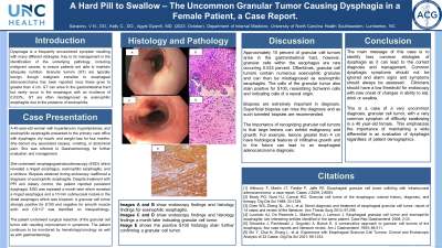
Introduction: Dysphagia is a relatively common symptom due to many different etiologies. Treating and identifying dysphagia is important to ensure patients are receiving adequate nutrition and to rule out potential underlying malignancies. Granular tumors are often benign, however, once greater than 4 cm can become esophageal adenocarcinoma. These can arise in the gastrointestinal tract, but rarely occur in the esophagus with an incidence of 0.033%, and are often misdiagnosed as eosinophilic esophagitis due to the presence of eosinophilia.
Case Description/Methods: A 40-year-old female with a past medical history of hypertension, hyperlipidemia, and eosinophilic esophagitis presented to the primary care office with difficulty swallowing, dry mouth, and weight loss for four months. She denied any associated nausea, vomiting, or abdominal pain. She was then referred to Gastroenterology for further management.
She received an EGD that showed a ringed esophagus, eosinophilic esophagitis, and a stricture. Biopsies were performed that further indicated eosinophilic esophagitis. However, the patient continued to have symptoms and an additional EGD was performed a month later. Repeat EGD showed a ringed esophagus and a 10 mm submucosal nodule in the distal esophagus that when biopsied showed a granular cell tumor strongly positive for S100 and negative for smooth muscle actin and CD117.
The patient underwent total resection of the granular cell tumor and had an improvement in symptoms. Presently, the patient is continued to be monitored by hematology and oncology as well as with Gastroenterology.
Discussion: Approximately 10 percent of granular cell tumors arise in the gastrointestinal tract, however, granular cells within the esophagus are rare and occur in about 0.033 percent. Oftentimes, granular cell tumors contain numerous eosinophilic granules and can then be misdiagnosed as eosinophilic esophagitis. The cells of the granular tumor also stain positive for S100, resembling Schwann cells and indicating cells of a neural origin.
Biopsies are extremely important in diagnosis. Superficial biopsies can miss the diagnosis and as such tunneled biopsies are recommended.
The importance of recognizing granular cell tumors is that large lesions can exhibit malignancy and growth. For example, lesions greater than 4 cm have histological features of infiltrative growth and in the future can lead to an esophageal adenocarcinoma diagnosis.

Disclosures:
Valentina N. Saracino, DO, Catherine Kelly, DO, Kwadwo Agyei-Gyamfi, MD. P0445 - A Hard Pill to Swallow - The Uncommon Granular Tumor Causing Dysphagia in a Female Patient: A Case Report, ACG 2023 Annual Scientific Meeting Abstracts. Vancouver, BC, Canada: American College of Gastroenterology.
Southeastern Regional Medical Center, Lumberton, NC
Introduction: Dysphagia is a relatively common symptom due to many different etiologies. Treating and identifying dysphagia is important to ensure patients are receiving adequate nutrition and to rule out potential underlying malignancies. Granular tumors are often benign, however, once greater than 4 cm can become esophageal adenocarcinoma. These can arise in the gastrointestinal tract, but rarely occur in the esophagus with an incidence of 0.033%, and are often misdiagnosed as eosinophilic esophagitis due to the presence of eosinophilia.
Case Description/Methods: A 40-year-old female with a past medical history of hypertension, hyperlipidemia, and eosinophilic esophagitis presented to the primary care office with difficulty swallowing, dry mouth, and weight loss for four months. She denied any associated nausea, vomiting, or abdominal pain. She was then referred to Gastroenterology for further management.
She received an EGD that showed a ringed esophagus, eosinophilic esophagitis, and a stricture. Biopsies were performed that further indicated eosinophilic esophagitis. However, the patient continued to have symptoms and an additional EGD was performed a month later. Repeat EGD showed a ringed esophagus and a 10 mm submucosal nodule in the distal esophagus that when biopsied showed a granular cell tumor strongly positive for S100 and negative for smooth muscle actin and CD117.
The patient underwent total resection of the granular cell tumor and had an improvement in symptoms. Presently, the patient is continued to be monitored by hematology and oncology as well as with Gastroenterology.
Discussion: Approximately 10 percent of granular cell tumors arise in the gastrointestinal tract, however, granular cells within the esophagus are rare and occur in about 0.033 percent. Oftentimes, granular cell tumors contain numerous eosinophilic granules and can then be misdiagnosed as eosinophilic esophagitis. The cells of the granular tumor also stain positive for S100, resembling Schwann cells and indicating cells of a neural origin.
Biopsies are extremely important in diagnosis. Superficial biopsies can miss the diagnosis and as such tunneled biopsies are recommended.
The importance of recognizing granular cell tumors is that large lesions can exhibit malignancy and growth. For example, lesions greater than 4 cm have histological features of infiltrative growth and in the future can lead to an esophageal adenocarcinoma diagnosis.

Figure: Images A and B show endoscopy and histology findings for eosinophilic esophagitis.
Images C and D taken a month later show endoscopy and histology findings indicating a granular cell tumor.
Image E shows the positive S100 histology stain further confirming a granular cell tumor.
Images C and D taken a month later show endoscopy and histology findings indicating a granular cell tumor.
Image E shows the positive S100 histology stain further confirming a granular cell tumor.
Disclosures:
Valentina Saracino indicated no relevant financial relationships.
Catherine Kelly indicated no relevant financial relationships.
Kwadwo Agyei-Gyamfi indicated no relevant financial relationships.
Valentina N. Saracino, DO, Catherine Kelly, DO, Kwadwo Agyei-Gyamfi, MD. P0445 - A Hard Pill to Swallow - The Uncommon Granular Tumor Causing Dysphagia in a Female Patient: A Case Report, ACG 2023 Annual Scientific Meeting Abstracts. Vancouver, BC, Canada: American College of Gastroenterology.
