Sunday Poster Session
Category: Esophagus
P0446 - A Purposeful Upper Endoscopy Exam Leading to Unexpected Findings
Sunday, October 22, 2023
3:30 PM - 7:00 PM PT
Location: Exhibit Hall

Has Audio

Sara Goff, MD
Temple University Hospital
Philadelphia, PA
Presenting Author(s)
Sara Goff, MD1, Daniel Baik, MD2, Woo Jung Lee, MBBCh3, Saraswathi Cappelle, DO1, Neena Mohan, MD1
1Temple University Hospital, Philadelphia, PA; 2Cooper University Health System, Mt. Laurel, NJ; 3Doylestown Health, Doyletown, NJ
Introduction: In this vignette we describe a patient who was found to have an unexpected subtle visible finding of esophageal squamous cell carcinoma during an upper endoscopy done for evaluation of epigastric pain and GERD. During this work-up she was then found to have a second mass in the presacral area of unclear significance with concern for a second malignancy. This case illustrates the importance of a thorough and diligent endoscopic exam. Endoscopy is an important diagnostic tool but remains provider dependent, with some studies noting over 11% of upper gastrointestinal neoplasms are missed on initial upper endoscopy.
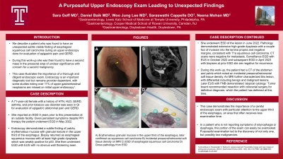
Case Description/Methods: A 71-year-old female with a history of HTN, HLD, GERD, asthma, and prior tobacco use disorder was seen in GI for evaluation of epigastric abdominal pain and GERD. She reported an EGD 5 years prior to this presentation at an outside facility. Given persistent symptoms despite PPI therapy the patient underwent EGD in May 2022. Endoscopy demonstrated a subtle finding of patchy erythematous mucosa with granular texture in the upper third of the esophagus. Biopsy returned as esophageal squamous mucosa with at least high grade dysplasia which was weakly positive for p53. She then underwent EGD with EUS with no obvious wall thickening seen. She underwent ESD of the lesion in June 2022. Pathology demonstrated extensive high-grade dysplasia with a couple foci of invasion into the lamina propria and negative margins, consistent with T1a squamous cell carcinoma. CT scans were negative for metastasis. She underwent surveillance EGD with EUS in October 2022 and subsequent EGD in April 2023 with biopsies at prior ESD site; these were negative for recurrence.
During this work-up, the patient had a CT of the abdomen and pelvis which noted an incidental presacral/retrorectal soft tissue density. An MRI further characterized this lesion, with differential including benign and malignant lesions. Later EUS with FNB demonstrated ‘atypical cytology.’ Tumor board recommended resection with colorectal surgery for definitive diagnosis, which the patient has deferred at this time.
Discussion: This case demonstrates the importance of a careful endoscopic exam with particular attention to the upper third of the esophagus, an area that often receives less examination time. In a patient who is not reporting symptoms of odynophagia or dysphagia, this portion of the exam can easily be overlooked. Purposeful examination led to not only the discovery of one, but possibly a second malignancy.

Disclosures:
Sara Goff, MD1, Daniel Baik, MD2, Woo Jung Lee, MBBCh3, Saraswathi Cappelle, DO1, Neena Mohan, MD1. P0446 - A Purposeful Upper Endoscopy Exam Leading to Unexpected Findings, ACG 2023 Annual Scientific Meeting Abstracts. Vancouver, BC, Canada: American College of Gastroenterology.
1Temple University Hospital, Philadelphia, PA; 2Cooper University Health System, Mt. Laurel, NJ; 3Doylestown Health, Doyletown, NJ
Introduction: In this vignette we describe a patient who was found to have an unexpected subtle visible finding of esophageal squamous cell carcinoma during an upper endoscopy done for evaluation of epigastric pain and GERD. During this work-up she was then found to have a second mass in the presacral area of unclear significance with concern for a second malignancy. This case illustrates the importance of a thorough and diligent endoscopic exam. Endoscopy is an important diagnostic tool but remains provider dependent, with some studies noting over 11% of upper gastrointestinal neoplasms are missed on initial upper endoscopy.
Case Description/Methods: A 71-year-old female with a history of HTN, HLD, GERD, asthma, and prior tobacco use disorder was seen in GI for evaluation of epigastric abdominal pain and GERD. She reported an EGD 5 years prior to this presentation at an outside facility. Given persistent symptoms despite PPI therapy the patient underwent EGD in May 2022. Endoscopy demonstrated a subtle finding of patchy erythematous mucosa with granular texture in the upper third of the esophagus. Biopsy returned as esophageal squamous mucosa with at least high grade dysplasia which was weakly positive for p53. She then underwent EGD with EUS with no obvious wall thickening seen. She underwent ESD of the lesion in June 2022. Pathology demonstrated extensive high-grade dysplasia with a couple foci of invasion into the lamina propria and negative margins, consistent with T1a squamous cell carcinoma. CT scans were negative for metastasis. She underwent surveillance EGD with EUS in October 2022 and subsequent EGD in April 2023 with biopsies at prior ESD site; these were negative for recurrence.
During this work-up, the patient had a CT of the abdomen and pelvis which noted an incidental presacral/retrorectal soft tissue density. An MRI further characterized this lesion, with differential including benign and malignant lesions. Later EUS with FNB demonstrated ‘atypical cytology.’ Tumor board recommended resection with colorectal surgery for definitive diagnosis, which the patient has deferred at this time.
Discussion: This case demonstrates the importance of a careful endoscopic exam with particular attention to the upper third of the esophagus, an area that often receives less examination time. In a patient who is not reporting symptoms of odynophagia or dysphagia, this portion of the exam can easily be overlooked. Purposeful examination led to not only the discovery of one, but possibly a second malignancy.

Figure: Figure 1. A) Erythematous granular mucosa in the upper third of the esophagus, later confirmed as squamous cell carcinoma B) Incidental presacral/retrorectal soft tissue density on MRI C) ESD of esophageal squamous cell carcinoma D) Gross pathology from ESD
Disclosures:
Sara Goff indicated no relevant financial relationships.
Daniel Baik indicated no relevant financial relationships.
Woo Jung Lee indicated no relevant financial relationships.
Saraswathi Cappelle indicated no relevant financial relationships.
Neena Mohan indicated no relevant financial relationships.
Sara Goff, MD1, Daniel Baik, MD2, Woo Jung Lee, MBBCh3, Saraswathi Cappelle, DO1, Neena Mohan, MD1. P0446 - A Purposeful Upper Endoscopy Exam Leading to Unexpected Findings, ACG 2023 Annual Scientific Meeting Abstracts. Vancouver, BC, Canada: American College of Gastroenterology.
