Sunday Poster Session
Category: Esophagus
P0447 - A Case of Esophagitis Dissecans Superficialis in a Patient Without Any Known Risk Factors
Sunday, October 22, 2023
3:30 PM - 7:00 PM PT
Location: Exhibit Hall

Has Audio

Rajdeepsingh Waghela, MD
Houston Methodist Hospital
Houston, TX
Presenting Author(s)
Rajdeepsingh Waghela, MD1, Shayan Amini, MD2, Sudha Kodali, MD, MPH1, David W. Victor, MD1, Tamneet Basra, MD1
1Houston Methodist Hospital, Houston, TX; 2Houston Methodist, Houston, TX
Introduction: Esophagitis dissecans superficialis (EDS) is a rare and benign condition of the esophagus characterized by sloughing of the esophageal mucosa. The endoscopic characteristics of EDS include whitish strips of peeling esophageal mucosa. While the etiology remains unknown, EDS is an extremely rare and benign condition and is often associated with autoimmune dermatological diseases, smoking, and certain medications. We report an incidental case of EDS in a patient with no history of pre-disposing known risk factors.
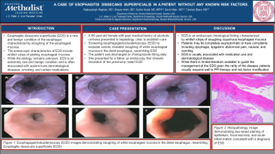
Case Description/Methods: A 66-year-old female with past medical history of alcoholic cirrhosis presented to hepatology clinic to establish care. Aside from intermittent constipation, the patient had no symptoms of GERD, dysphagia, or any denied any other complaints. Physical examination was grossly unremarkable. Lab work was significant for creatinine of 3.08 and ammonia level of 114. MRI abdomen was significant for cirrhosis with portal hypertension and evidence of para-esophageal varices. Her medications included spironolactone, furosemide, and propranolol. Screening esophagogastroduodenoscopy (EGD) to evaluate varices revealed sloughing of white esophageal mucosa in the distal esophagus, resembling ESD. Biopsies were obtained and pathologic examination of the sample showed two-toned staining of epithelium, focal necrosis, and acute inflammation consistent with a diagnosis of ESD. The patient was discharged on Pantoprazole 40mg daily. She presented for a follow up endoscopy that showed resolution of her previously noted EGD.
Discussion: EDS is an endoscopic-histological finding characterized by whitish strips of sloughing squamous esophageal mucosa. Histopathology evaluation of biopsy samples is notable for a constellation of non-specific findings including splitting of squamous epithelium, variably sized cysts and bullae, basal cell hyperplasia, parakeratosis, and focal or minimal inflammation. Patients may be completely asymptomatic or have complaints including dysphagia, epigastric abdominal pain, nausea, and vomiting. While EDS is usually associated with medication use and dermatological disease, our patient did not have any known pre-disposing risk factors. While there is limited literature available to guide the management of the EDS given the rarity of the disease, patients usually respond well to PPI therapy and risk factor modification. Our patient showed complete resolution of EDS after successful treatment with PPI therapy.

Disclosures:
Rajdeepsingh Waghela, MD1, Shayan Amini, MD2, Sudha Kodali, MD, MPH1, David W. Victor, MD1, Tamneet Basra, MD1. P0447 - A Case of Esophagitis Dissecans Superficialis in a Patient Without Any Known Risk Factors, ACG 2023 Annual Scientific Meeting Abstracts. Vancouver, BC, Canada: American College of Gastroenterology.
1Houston Methodist Hospital, Houston, TX; 2Houston Methodist, Houston, TX
Introduction: Esophagitis dissecans superficialis (EDS) is a rare and benign condition of the esophagus characterized by sloughing of the esophageal mucosa. The endoscopic characteristics of EDS include whitish strips of peeling esophageal mucosa. While the etiology remains unknown, EDS is an extremely rare and benign condition and is often associated with autoimmune dermatological diseases, smoking, and certain medications. We report an incidental case of EDS in a patient with no history of pre-disposing known risk factors.
Case Description/Methods: A 66-year-old female with past medical history of alcoholic cirrhosis presented to hepatology clinic to establish care. Aside from intermittent constipation, the patient had no symptoms of GERD, dysphagia, or any denied any other complaints. Physical examination was grossly unremarkable. Lab work was significant for creatinine of 3.08 and ammonia level of 114. MRI abdomen was significant for cirrhosis with portal hypertension and evidence of para-esophageal varices. Her medications included spironolactone, furosemide, and propranolol. Screening esophagogastroduodenoscopy (EGD) to evaluate varices revealed sloughing of white esophageal mucosa in the distal esophagus, resembling ESD. Biopsies were obtained and pathologic examination of the sample showed two-toned staining of epithelium, focal necrosis, and acute inflammation consistent with a diagnosis of ESD. The patient was discharged on Pantoprazole 40mg daily. She presented for a follow up endoscopy that showed resolution of her previously noted EGD.
Discussion: EDS is an endoscopic-histological finding characterized by whitish strips of sloughing squamous esophageal mucosa. Histopathology evaluation of biopsy samples is notable for a constellation of non-specific findings including splitting of squamous epithelium, variably sized cysts and bullae, basal cell hyperplasia, parakeratosis, and focal or minimal inflammation. Patients may be completely asymptomatic or have complaints including dysphagia, epigastric abdominal pain, nausea, and vomiting. While EDS is usually associated with medication use and dermatological disease, our patient did not have any known pre-disposing risk factors. While there is limited literature available to guide the management of the EDS given the rarity of the disease, patients usually respond well to PPI therapy and risk factor modification. Our patient showed complete resolution of EDS after successful treatment with PPI therapy.

Figure: (A), (B), (C) Visualized Appearance of Lower Third of Esophagus on Initial EGD; (D), (E), (F) Visualized Appearance of Lower Third of Esophagus on Follow-Up EGD
Disclosures:
Rajdeepsingh Waghela indicated no relevant financial relationships.
Shayan Amini indicated no relevant financial relationships.
Sudha Kodali indicated no relevant financial relationships.
David Victor indicated no relevant financial relationships.
Tamneet Basra indicated no relevant financial relationships.
Rajdeepsingh Waghela, MD1, Shayan Amini, MD2, Sudha Kodali, MD, MPH1, David W. Victor, MD1, Tamneet Basra, MD1. P0447 - A Case of Esophagitis Dissecans Superficialis in a Patient Without Any Known Risk Factors, ACG 2023 Annual Scientific Meeting Abstracts. Vancouver, BC, Canada: American College of Gastroenterology.
