Sunday Poster Session
Category: Esophagus
P0450 - Parakeratosis of the Esophagus: A Rare Mimic of Eosinophilic Esophagitis
Sunday, October 22, 2023
3:30 PM - 7:00 PM PT
Location: Exhibit Hall

Has Audio
- SP
Sohum A. Patwa, MD
Warren Alpert Medical School of Brown University
Providence, RI
Presenting Author(s)
Sohum A. Patwa, MD1, John Paul Nsubuga, MD2, Amer Malik, MD3
1Warren Alpert Medical School of Brown University, Providence, RI; 2Brown University, Providence, RI; 3Lifespan, Providence, RI
Introduction: Parakeratosis is a histologic finding resulting from abnormal maturation of epithelial keratinocytes, sometimes seen in inflammatory skin disorders such as psoriasis. Parakeratosis of the esophagus is a rarely reported pathologic finding. Some reported cases attribute its development to vitamin or mineral deficiencies, though the definite etiology remains unknown. Its reported endoscopic appearance has varied. To our knowledge, a case of esophageal parakeratosis mimicking eosinophilic esophagitis has not been reported. We report the rare case of a presentation and endoscopic appearance mimicking that of eosinophilic esophagitis but with biopsies consistent with parakeratosis of the esophagus.
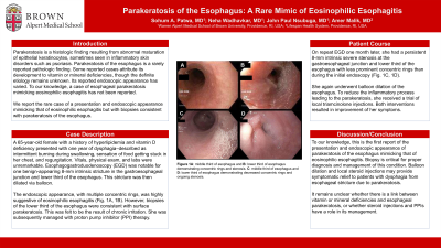
Case Description/Methods: A 65-year-old female with a history of hyperlipidemia and vitamin D deficiency presented with one year of dysphagia – described as intermittent burning during swallowing, sensation of food getting stuck in her chest, and regurgitation. Vitals, physical exam, and labs were unremarkable. Esophagogastroduodenoscopy (EGD) was notable for one benign-appearing 8-mm intrinsic stricture in the gastroesophageal junction and lower third of the esophagus. This stricture was then dilated via balloon. The endoscopic appearance, with multiple concentric rings, was highly suggestive of eosinophilic esophagitis (Fig. 1A, 1B). However, biopsies of the lower third of the esophagus were consistent with surface parakeratosis. This was felt to be the result of chronic irritation. She was subsequently managed with proton pump inhibitor (PPI) therapy.
On repeat EGD one month later, she had a persistent 8-mm intrinsic severe stenosis at the gastroesophageal junction and lower third of the esophagus with less prominent concentric rings than during the initial endoscopy (Fig. 1C, 1D). She again underwent balloon dilation of the esophagus. She was planned to trial triamcinolone injections to reduce the inflammatory process leading to the parakeratosis.
Discussion: To our knowledge, this is the first report of the presentation and endoscopic appearance of parakeratosis of the esophagus mimicking that of eosinophilic esophagitis. Biopsy is critical for proper diagnosis and management of this condition. Balloon dilation may provide symptomatic relief to patients with dysphagia due to esophageal stricture from parakeratosis. It remains unclear whether there is a link between vitamin or mineral deficiencies and esophageal parakeratosis, or whether steroid injections and PPIs have a role in its management.

Disclosures:
Sohum A. Patwa, MD1, John Paul Nsubuga, MD2, Amer Malik, MD3. P0450 - Parakeratosis of the Esophagus: A Rare Mimic of Eosinophilic Esophagitis, ACG 2023 Annual Scientific Meeting Abstracts. Vancouver, BC, Canada: American College of Gastroenterology.
1Warren Alpert Medical School of Brown University, Providence, RI; 2Brown University, Providence, RI; 3Lifespan, Providence, RI
Introduction: Parakeratosis is a histologic finding resulting from abnormal maturation of epithelial keratinocytes, sometimes seen in inflammatory skin disorders such as psoriasis. Parakeratosis of the esophagus is a rarely reported pathologic finding. Some reported cases attribute its development to vitamin or mineral deficiencies, though the definite etiology remains unknown. Its reported endoscopic appearance has varied. To our knowledge, a case of esophageal parakeratosis mimicking eosinophilic esophagitis has not been reported. We report the rare case of a presentation and endoscopic appearance mimicking that of eosinophilic esophagitis but with biopsies consistent with parakeratosis of the esophagus.
Case Description/Methods: A 65-year-old female with a history of hyperlipidemia and vitamin D deficiency presented with one year of dysphagia – described as intermittent burning during swallowing, sensation of food getting stuck in her chest, and regurgitation. Vitals, physical exam, and labs were unremarkable. Esophagogastroduodenoscopy (EGD) was notable for one benign-appearing 8-mm intrinsic stricture in the gastroesophageal junction and lower third of the esophagus. This stricture was then dilated via balloon. The endoscopic appearance, with multiple concentric rings, was highly suggestive of eosinophilic esophagitis (Fig. 1A, 1B). However, biopsies of the lower third of the esophagus were consistent with surface parakeratosis. This was felt to be the result of chronic irritation. She was subsequently managed with proton pump inhibitor (PPI) therapy.
On repeat EGD one month later, she had a persistent 8-mm intrinsic severe stenosis at the gastroesophageal junction and lower third of the esophagus with less prominent concentric rings than during the initial endoscopy (Fig. 1C, 1D). She again underwent balloon dilation of the esophagus. She was planned to trial triamcinolone injections to reduce the inflammatory process leading to the parakeratosis.
Discussion: To our knowledge, this is the first report of the presentation and endoscopic appearance of parakeratosis of the esophagus mimicking that of eosinophilic esophagitis. Biopsy is critical for proper diagnosis and management of this condition. Balloon dilation may provide symptomatic relief to patients with dysphagia due to esophageal stricture from parakeratosis. It remains unclear whether there is a link between vitamin or mineral deficiencies and esophageal parakeratosis, or whether steroid injections and PPIs have a role in its management.

Figure: A: middle third of esophagus and B: lower third of esophagus demonstrating concentric rings and stenosis. C: middle third of esophagus and D: lower third of esophagus one month later demonstrating decreased concentric rings and ongoing stenosis.
Disclosures:
Sohum Patwa indicated no relevant financial relationships.
John Paul Nsubuga indicated no relevant financial relationships.
Amer Malik indicated no relevant financial relationships.
Sohum A. Patwa, MD1, John Paul Nsubuga, MD2, Amer Malik, MD3. P0450 - Parakeratosis of the Esophagus: A Rare Mimic of Eosinophilic Esophagitis, ACG 2023 Annual Scientific Meeting Abstracts. Vancouver, BC, Canada: American College of Gastroenterology.
