Sunday Poster Session
Category: Esophagus
P0457 - Boerhaave Syndrome Mimicking Acute Coronary Syndrome
Sunday, October 22, 2023
3:30 PM - 7:00 PM PT
Location: Exhibit Hall

Has Audio

Reid Schalet, DO
George Washington University Hospital
Washington, DC
Presenting Author(s)
Award: Presidential Poster Award
Reid Schalet, DO1, Francis Carro, MD1, Cyrus Adams-Mardi, MD, MS2, Hanna Haile, BS1, Samuel A. Schueler, MD1, Marie L. Borum, MD, EdD, MPH3
1George Washington University Hospital, Washington, DC; 2George Washington University School of Medicine, Frederick, MD; 3George Washington University Medical Center, Washington, DC
Introduction: Boerhaave syndrome, a spontaneous esophageal perforation in the setting of increased intraesophageal pressure, is associated with significant morbidity and mortality. It is a rare condition with an estimated annual incidence of 3.1 /1,000,000. This is an unusual case of Boerhaave syndrome with associated EKG and cardiac enzymes abnormalities prompting a diagnosis of acute coronary syndrome.
Case Description/Methods: A 63-year-old man with HIV, maintained on highly active antiretroviral therapy (CD4 counts in 400s), prior pulmonary embolism (not on anticoagulation) presented to the ER with 3 days of persistent, diffuse chest pain radiating to his abdomen, dyspnea and fatigue. Exam revealed a pale-appearing male who was hypotensive and had chest wall and abdominal tenderness with involuntary guarding. An EKG showed inferolateral ST elevations. Labs demonstrated a bandemia of 19%, lactate of 4.9 and troponin of 0.013. During a coronary angiography, a drug-eluting stent was placed in a 70% occlusion of the left anterior descending artery (LAD).
Following angiography, he had continued chest and abdominal discomfort with ST elevations on EKG. CT chest angiography showed an esophageal perforation at the level of the carina, diffuse pneumomediastinum and possible mediastinitis. Contrast extravasation on esophagram confirmed perforation. An EGD showed nodular, necrotic and friable esophageal mucosa with 4 large perforations occurring 25-40 cm from the incisors. Fluoroscopy-guided placement of fully covered flexible stents were performed. He remained intubated for 2 weeks until end of life.
Medical records noted that 5 months prior, a neck CT scan performed for dysphagia, revealed upper esophageal distension with air and fluid layering in the mid-esophagus at the level of the carina associated with a lobulated wall thickening consistent with possible neoplasm. He left against medical advice.
Discussion: Boerhaave syndrome typically presents with severe chest, neck or upper abdominal pain and subcutaneous emphysema often associated with vomiting or retching, although atypical presentations may occur. Esophageal perforation may result in pericardial irritation due to mediastinal proximity which can mimic acute coronary syndrome. This case was notable for persistent symptoms and EKG changes despite LAD stenting of a 70% occlusion. It is critical that there is a high index of suspicion for Boerhaave syndrome in appropriate clinical circumstances to potentially improve clinical outcomes.

Disclosures:
Reid Schalet, DO1, Francis Carro, MD1, Cyrus Adams-Mardi, MD, MS2, Hanna Haile, BS1, Samuel A. Schueler, MD1, Marie L. Borum, MD, EdD, MPH3. P0457 - Boerhaave Syndrome Mimicking Acute Coronary Syndrome, ACG 2023 Annual Scientific Meeting Abstracts. Vancouver, BC, Canada: American College of Gastroenterology.
Reid Schalet, DO1, Francis Carro, MD1, Cyrus Adams-Mardi, MD, MS2, Hanna Haile, BS1, Samuel A. Schueler, MD1, Marie L. Borum, MD, EdD, MPH3
1George Washington University Hospital, Washington, DC; 2George Washington University School of Medicine, Frederick, MD; 3George Washington University Medical Center, Washington, DC
Introduction: Boerhaave syndrome, a spontaneous esophageal perforation in the setting of increased intraesophageal pressure, is associated with significant morbidity and mortality. It is a rare condition with an estimated annual incidence of 3.1 /1,000,000. This is an unusual case of Boerhaave syndrome with associated EKG and cardiac enzymes abnormalities prompting a diagnosis of acute coronary syndrome.
Case Description/Methods: A 63-year-old man with HIV, maintained on highly active antiretroviral therapy (CD4 counts in 400s), prior pulmonary embolism (not on anticoagulation) presented to the ER with 3 days of persistent, diffuse chest pain radiating to his abdomen, dyspnea and fatigue. Exam revealed a pale-appearing male who was hypotensive and had chest wall and abdominal tenderness with involuntary guarding. An EKG showed inferolateral ST elevations. Labs demonstrated a bandemia of 19%, lactate of 4.9 and troponin of 0.013. During a coronary angiography, a drug-eluting stent was placed in a 70% occlusion of the left anterior descending artery (LAD).
Following angiography, he had continued chest and abdominal discomfort with ST elevations on EKG. CT chest angiography showed an esophageal perforation at the level of the carina, diffuse pneumomediastinum and possible mediastinitis. Contrast extravasation on esophagram confirmed perforation. An EGD showed nodular, necrotic and friable esophageal mucosa with 4 large perforations occurring 25-40 cm from the incisors. Fluoroscopy-guided placement of fully covered flexible stents were performed. He remained intubated for 2 weeks until end of life.
Medical records noted that 5 months prior, a neck CT scan performed for dysphagia, revealed upper esophageal distension with air and fluid layering in the mid-esophagus at the level of the carina associated with a lobulated wall thickening consistent with possible neoplasm. He left against medical advice.
Discussion: Boerhaave syndrome typically presents with severe chest, neck or upper abdominal pain and subcutaneous emphysema often associated with vomiting or retching, although atypical presentations may occur. Esophageal perforation may result in pericardial irritation due to mediastinal proximity which can mimic acute coronary syndrome. This case was notable for persistent symptoms and EKG changes despite LAD stenting of a 70% occlusion. It is critical that there is a high index of suspicion for Boerhaave syndrome in appropriate clinical circumstances to potentially improve clinical outcomes.

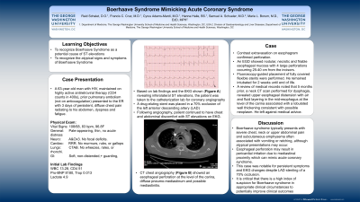
Figure: 1A. EKG showing anterolateral and inferior ST elevations. 1B. CT Angiogram with findings of an esophageal perforation at the level of the carina, diffuse pneumomediastinum and possible mediastinitis
Disclosures:
Reid Schalet indicated no relevant financial relationships.
Francis Carro indicated no relevant financial relationships.
Cyrus Adams-Mardi indicated no relevant financial relationships.
Hanna Haile indicated no relevant financial relationships.
Samuel Schueler indicated no relevant financial relationships.
Marie Borum: Takeda – Advisory Committee/Board Member, Speakers Bureau.
Reid Schalet, DO1, Francis Carro, MD1, Cyrus Adams-Mardi, MD, MS2, Hanna Haile, BS1, Samuel A. Schueler, MD1, Marie L. Borum, MD, EdD, MPH3. P0457 - Boerhaave Syndrome Mimicking Acute Coronary Syndrome, ACG 2023 Annual Scientific Meeting Abstracts. Vancouver, BC, Canada: American College of Gastroenterology.

