Sunday Poster Session
Category: Esophagus
P0459 - Esophagitis Dissecans Superficialis: When the Esophagus Sheds Its Skin
Sunday, October 22, 2023
3:30 PM - 7:00 PM PT
Location: Exhibit Hall

Has Audio
.jpg)
Joseph David, MD
University of Arizona College of Medicine
Phoenix, AZ
Presenting Author(s)
Brian M.. Fung, MD, Joseph David, MD
University of Arizona College of Medicine, Phoenix, AZ
Introduction: Esophagitis dissecans superficialis is a rare, benign endoscopic finding characterized by sloughing of superficial esophageal mucosa. The finding has an especially unique appearance, with endoscopy frequently revealing sheets of peeling superficial mucosa and a normal underlying basal mucosa. Herein, we present a case of esophagitis dissecans superficialis found on diagnostic upper endoscopy for an unrelated reason.
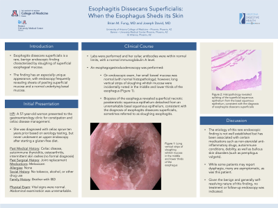
Case Description/Methods: A 57-year-old woman with a history of celiac disease, autoimmune thyroiditis, osteoarthritis, and intermittent skin rashes without formal diagnosis presented to the gastroenterology clinic for constipation and celiac disease management. She was diagnosed with celiac sprue ten years prior based on serology testing, but never underwent an upper endoscopy after starting a gluten-free diet. Labs were performed and her celiac antibodies were within normal limits, with a normal immunoglobulin A level. An esophagogastroduodenoscopy was performed. On exam, her small bowel mucosa was normal (with normal histopathology); however, long vertical strips of sloughing whitish mucosa were incidentally noted in the middle and lower thirds of the esophagus (Figure 1). Biopsies of the esophagus revealed superficial necrotic parakeratotic squamous epithelium detached from an unremarkable basal squamous epithelium, consistent with the diagnosis of esophagitis dissecans superficialis, sometimes referred to as sloughing esophagitis.
Discussion: The etiology of this rare endoscopic finding is not well established but has been associated with certain medications such as non-steroidal anti-inflammatory drugs (which this patient was taking for her arthritis), autoimmune conditions (such as celiac disease), debility, as well as bullous skin disorders (such as pemphigus vulgaris). While some patients may report dysphagia, many are asymptomatic, as was this patient. Given the benign and generally self-resolving nature of this finding, no treatment or follow-up endoscopy was indicated.

Disclosures:
Brian M.. Fung, MD, Joseph David, MD. P0459 - Esophagitis Dissecans Superficialis: When the Esophagus Sheds Its Skin, ACG 2023 Annual Scientific Meeting Abstracts. Vancouver, BC, Canada: American College of Gastroenterology.
University of Arizona College of Medicine, Phoenix, AZ
Introduction: Esophagitis dissecans superficialis is a rare, benign endoscopic finding characterized by sloughing of superficial esophageal mucosa. The finding has an especially unique appearance, with endoscopy frequently revealing sheets of peeling superficial mucosa and a normal underlying basal mucosa. Herein, we present a case of esophagitis dissecans superficialis found on diagnostic upper endoscopy for an unrelated reason.
Case Description/Methods: A 57-year-old woman with a history of celiac disease, autoimmune thyroiditis, osteoarthritis, and intermittent skin rashes without formal diagnosis presented to the gastroenterology clinic for constipation and celiac disease management. She was diagnosed with celiac sprue ten years prior based on serology testing, but never underwent an upper endoscopy after starting a gluten-free diet. Labs were performed and her celiac antibodies were within normal limits, with a normal immunoglobulin A level. An esophagogastroduodenoscopy was performed. On exam, her small bowel mucosa was normal (with normal histopathology); however, long vertical strips of sloughing whitish mucosa were incidentally noted in the middle and lower thirds of the esophagus (Figure 1). Biopsies of the esophagus revealed superficial necrotic parakeratotic squamous epithelium detached from an unremarkable basal squamous epithelium, consistent with the diagnosis of esophagitis dissecans superficialis, sometimes referred to as sloughing esophagitis.
Discussion: The etiology of this rare endoscopic finding is not well established but has been associated with certain medications such as non-steroidal anti-inflammatory drugs (which this patient was taking for her arthritis), autoimmune conditions (such as celiac disease), debility, as well as bullous skin disorders (such as pemphigus vulgaris). While some patients may report dysphagia, many are asymptomatic, as was this patient. Given the benign and generally self-resolving nature of this finding, no treatment or follow-up endoscopy was indicated.

Figure: Vertical strips of sloughing whitish mucosa in the mid and distal esophagus, consistent with esophagitis dissecans superficialis.
Disclosures:
Brian Fung indicated no relevant financial relationships.
Joseph David indicated no relevant financial relationships.
Brian M.. Fung, MD, Joseph David, MD. P0459 - Esophagitis Dissecans Superficialis: When the Esophagus Sheds Its Skin, ACG 2023 Annual Scientific Meeting Abstracts. Vancouver, BC, Canada: American College of Gastroenterology.
