Sunday Poster Session
Category: Esophagus
P0461 - Esophageal Parakeratosis: A Rare Finding During Evaluation of Abdominal Pain
Sunday, October 22, 2023
3:30 PM - 7:00 PM PT
Location: Exhibit Hall

Has Audio
- TG
Tyler Grantham, MD
Staten Island University Hospital
Staten Island, NY
Presenting Author(s)
Rajarajeshwari Ramachandran, MD1, Tyler Grantham, MD2, Vikash Kumar, MD1, Heidi Budke, MD3, Vinaya Gaduputi, MD3
1Brooklyn Hospital Center, Brooklyn, NY; 2Staten Island University Hospital, Staten Island, NY; 3Blanchard Valley Health System, Findlay, OH
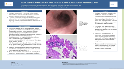
Introduction: Esophageal parakeratosis is a rare, asymptomatic benign finding that is characterized by the presence of incomplete keratinized squamous epithelium in the esophagus. This is the case of a 61-year-old man who underwent esophagogastroduodenoscopy (EGD) for the evaluation of chronic abdominal pain and was incidentally diagnosed with esophageal parakeratosis.
Case Description/Methods: A 61-year-old man with anxiety disorder and benign prostate hypertrophy presented for evaluation for chronic upper abdominal pain associated with nausea for several months. He denied dysphagia, odynophagia, vomiting, hematemesis, diarrhea, constipation, melena, hematochezia, and unintentional weight loss. He endorsed current intermittent marijuana use. Physical examination was negative for abdominal tenderness, signs of bowel obstruction, or palpable mass. Laboratory data: hemoglobin 15.9 g/dL with normal electrolytes, glucose, and liver enzymes. Patient underwent EGD which revealed scattered, small, whitish mucosal plaques in the mid and lower third of the esophagus (image A) and rest of the examination was unremarkable. Biopsies of the esophageal lesions demonstrated prominence of the basilar zone, and superficial parakeratotic debris consistent with esophageal parakeratosis (image B). He was advised to abstain from marijuana use and during his 6 month follow up had resolution of abdominal pain and nausea.
Discussion: Esophageal parakeratosis presents as white, membranous, linear plaques and flat elevated lesions. It is has been associated with zinc deficiency, B12 deficiency, tyolysis, ethanol use, and smoking. The histopathological distinction of this benign lesion carries significance as gross appearance can raise concern for neoplastic lesions. Reports have shown the presence of parakeratosis in patients with esophageal cancer, however, there is no evidence that parakeratosis progresses to dysplasia or cancer. There are currently no recommendations on treatment or surveillance of esophageal parakeratosis. Our patient was incidentally diagnosed with esophageal parakeratosis while undergoing evaluation of nausea and upper abdominal pain which resolved after cessation of marijuana use.

Disclosures:
Rajarajeshwari Ramachandran, MD1, Tyler Grantham, MD2, Vikash Kumar, MD1, Heidi Budke, MD3, Vinaya Gaduputi, MD3. P0461 - Esophageal Parakeratosis: A Rare Finding During Evaluation of Abdominal Pain, ACG 2023 Annual Scientific Meeting Abstracts. Vancouver, BC, Canada: American College of Gastroenterology.
1Brooklyn Hospital Center, Brooklyn, NY; 2Staten Island University Hospital, Staten Island, NY; 3Blanchard Valley Health System, Findlay, OH
Introduction: Esophageal parakeratosis is a rare, asymptomatic benign finding that is characterized by the presence of incomplete keratinized squamous epithelium in the esophagus. This is the case of a 61-year-old man who underwent esophagogastroduodenoscopy (EGD) for the evaluation of chronic abdominal pain and was incidentally diagnosed with esophageal parakeratosis.
Case Description/Methods: A 61-year-old man with anxiety disorder and benign prostate hypertrophy presented for evaluation for chronic upper abdominal pain associated with nausea for several months. He denied dysphagia, odynophagia, vomiting, hematemesis, diarrhea, constipation, melena, hematochezia, and unintentional weight loss. He endorsed current intermittent marijuana use. Physical examination was negative for abdominal tenderness, signs of bowel obstruction, or palpable mass. Laboratory data: hemoglobin 15.9 g/dL with normal electrolytes, glucose, and liver enzymes. Patient underwent EGD which revealed scattered, small, whitish mucosal plaques in the mid and lower third of the esophagus (image A) and rest of the examination was unremarkable. Biopsies of the esophageal lesions demonstrated prominence of the basilar zone, and superficial parakeratotic debris consistent with esophageal parakeratosis (image B). He was advised to abstain from marijuana use and during his 6 month follow up had resolution of abdominal pain and nausea.
Discussion: Esophageal parakeratosis presents as white, membranous, linear plaques and flat elevated lesions. It is has been associated with zinc deficiency, B12 deficiency, tyolysis, ethanol use, and smoking. The histopathological distinction of this benign lesion carries significance as gross appearance can raise concern for neoplastic lesions. Reports have shown the presence of parakeratosis in patients with esophageal cancer, however, there is no evidence that parakeratosis progresses to dysplasia or cancer. There are currently no recommendations on treatment or surveillance of esophageal parakeratosis. Our patient was incidentally diagnosed with esophageal parakeratosis while undergoing evaluation of nausea and upper abdominal pain which resolved after cessation of marijuana use.

Figure: Image A: EGD image - scattered whitish mucosal plaques in the esophagus (red arrow)
Image B: Histopathology image - prominence of the basilar zone, and superficial parakeratotic debris (black arrow)
Image B: Histopathology image - prominence of the basilar zone, and superficial parakeratotic debris (black arrow)
Disclosures:
Rajarajeshwari Ramachandran indicated no relevant financial relationships.
Tyler Grantham indicated no relevant financial relationships.
Vikash Kumar indicated no relevant financial relationships.
Heidi Budke indicated no relevant financial relationships.
Vinaya Gaduputi indicated no relevant financial relationships.
Rajarajeshwari Ramachandran, MD1, Tyler Grantham, MD2, Vikash Kumar, MD1, Heidi Budke, MD3, Vinaya Gaduputi, MD3. P0461 - Esophageal Parakeratosis: A Rare Finding During Evaluation of Abdominal Pain, ACG 2023 Annual Scientific Meeting Abstracts. Vancouver, BC, Canada: American College of Gastroenterology.
