Sunday Poster Session
Category: Esophagus
P0475 - A Stained Esophagus: A Rare Case of Esophageal Melanocytosis
Sunday, October 22, 2023
3:30 PM - 7:00 PM PT
Location: Exhibit Hall

Has Audio

Aboud Kaliounji, MD
SUNY Downstate Health Sciences University
Brooklyn, NY
Presenting Author(s)
Aboud Kaliounji, MD1, Vimal Bodiwala, MD1, Qi Yu, MD, MPH2, Eric Steimetz, MD1, Zhongua Li, MD1
1SUNY Downstate Health Sciences University, Brooklyn, NY; 2SUNY Downstate Health Sciences University/NYCHHC Kings County Hospital, Brooklyn, NY
Introduction: Esophageal melanocytosis is a rare benign pathology of the gastrointestinal tract characterized by accumulation of melanin pigment in the esophageal mucosa and melanocytic proliferation in the basal layer of the squamous epithelium. It is a benign condition that has only been only reported a few dozen times, mainly in India and Japan and less commonly in Western countries, with an incidence ranging between 0.07 and 2.1% in a consecutive endoscopy series. Here, we report a case of esophageal melanocytosis found incidentally in a woman being evaluated for unintentional weight loss.
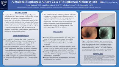
Case Description/Methods: A 70-year-old African-American female with a past medical history of hypertension, type 2 diabetes, right breast cancer status post lumpectomy and chemoradiotherapy, heart failure was referred to the gastroenterology service for evaluation of unintentional weight loss. The patient endorsed 30-pound weight loss, dyspepsia, early satiety and decreased appetite over the past year. She denied any nausea, vomiting, diarrhea, abdominal pain, hematochezia, melena, acid reflux, or dysphagia. Physical exam and laboratory work, including fecal occult blood test, were unremarkable. Computed Tomography abdomen/pelvis with contrast was negative for neoplasm and lymphadenopathy. Colonoscopy revealed diverticulosis and a 5 mm hyperplastic polyp. Esophagogastroduodenoscopy (EGD) showed diffuse mild mucosal discoloration of the entire esophagus and erythematous and eroded gastric mucosa (Figure 1). Mid and distal esophageal biopsies revealed benign squamous epithelium with changes consistent with esophageal melanocytosis (Figure 1). Immunohistochemical stains for melanocytic markers, melan A and SOX10, highlighted the benign melanocytic proliferation. She was sheduled for closed follow-up and repeat EGD for surveillance.
Discussion: Due to its rarity in the gastrointestinal tract, little is known about the etiology and natural course of this condition. It has been reported more in males (2:1 ratio) and is commonly found in the middle and lower third of the esophagus. It appears to be associated with chronic esophageal stimuli such as acid reflux, smoking and alcohol and was also found in rare systemic diseases such as Addison’s and Celiac. Although mainly asymptomatic, it has been suggested to be a precursor of primary esophageal melanoma and highlights the importance of our finding. Due to its low prevalence, no managment or surveillance guidelines have been published.

Disclosures:
Aboud Kaliounji, MD1, Vimal Bodiwala, MD1, Qi Yu, MD, MPH2, Eric Steimetz, MD1, Zhongua Li, MD1. P0475 - A Stained Esophagus: A Rare Case of Esophageal Melanocytosis, ACG 2023 Annual Scientific Meeting Abstracts. Vancouver, BC, Canada: American College of Gastroenterology.
1SUNY Downstate Health Sciences University, Brooklyn, NY; 2SUNY Downstate Health Sciences University/NYCHHC Kings County Hospital, Brooklyn, NY
Introduction: Esophageal melanocytosis is a rare benign pathology of the gastrointestinal tract characterized by accumulation of melanin pigment in the esophageal mucosa and melanocytic proliferation in the basal layer of the squamous epithelium. It is a benign condition that has only been only reported a few dozen times, mainly in India and Japan and less commonly in Western countries, with an incidence ranging between 0.07 and 2.1% in a consecutive endoscopy series. Here, we report a case of esophageal melanocytosis found incidentally in a woman being evaluated for unintentional weight loss.
Case Description/Methods: A 70-year-old African-American female with a past medical history of hypertension, type 2 diabetes, right breast cancer status post lumpectomy and chemoradiotherapy, heart failure was referred to the gastroenterology service for evaluation of unintentional weight loss. The patient endorsed 30-pound weight loss, dyspepsia, early satiety and decreased appetite over the past year. She denied any nausea, vomiting, diarrhea, abdominal pain, hematochezia, melena, acid reflux, or dysphagia. Physical exam and laboratory work, including fecal occult blood test, were unremarkable. Computed Tomography abdomen/pelvis with contrast was negative for neoplasm and lymphadenopathy. Colonoscopy revealed diverticulosis and a 5 mm hyperplastic polyp. Esophagogastroduodenoscopy (EGD) showed diffuse mild mucosal discoloration of the entire esophagus and erythematous and eroded gastric mucosa (Figure 1). Mid and distal esophageal biopsies revealed benign squamous epithelium with changes consistent with esophageal melanocytosis (Figure 1). Immunohistochemical stains for melanocytic markers, melan A and SOX10, highlighted the benign melanocytic proliferation. She was sheduled for closed follow-up and repeat EGD for surveillance.
Discussion: Due to its rarity in the gastrointestinal tract, little is known about the etiology and natural course of this condition. It has been reported more in males (2:1 ratio) and is commonly found in the middle and lower third of the esophagus. It appears to be associated with chronic esophageal stimuli such as acid reflux, smoking and alcohol and was also found in rare systemic diseases such as Addison’s and Celiac. Although mainly asymptomatic, it has been suggested to be a precursor of primary esophageal melanoma and highlights the importance of our finding. Due to its low prevalence, no managment or surveillance guidelines have been published.

Figure: Figure 1: A) Esophageal biopsy showing an increased number of melanocytes in the basal layer of esophageal squamous epithelium and an increased quantity of melanin in the esophageal mucosa (H&E, x 200). B) Biopsy showing melanocytes highlighted by melanocytic markers SOX10 (x 200). C) EGD white light image showing discoloration of the mucosa throughout the esophagus. D) EGD narrow-band image showing discoloration of the mucosa.
Disclosures:
Aboud Kaliounji indicated no relevant financial relationships.
Vimal Bodiwala indicated no relevant financial relationships.
Qi Yu indicated no relevant financial relationships.
Eric Steimetz indicated no relevant financial relationships.
Zhongua Li indicated no relevant financial relationships.
Aboud Kaliounji, MD1, Vimal Bodiwala, MD1, Qi Yu, MD, MPH2, Eric Steimetz, MD1, Zhongua Li, MD1. P0475 - A Stained Esophagus: A Rare Case of Esophageal Melanocytosis, ACG 2023 Annual Scientific Meeting Abstracts. Vancouver, BC, Canada: American College of Gastroenterology.
