Sunday Poster Session
Category: Esophagus
P0503 - Long Strange Trip: The Story of a Migrating Ballistic Fragment
Sunday, October 22, 2023
3:30 PM - 7:00 PM PT
Location: Exhibit Hall

Has Audio

Matthew Houle, MD
Tulane University School of Medicine
New Orleans, LA
Presenting Author(s)
Matthew Houle, MD, John Bloch, MD, MPH, Kaitlin Occhipinti, MD
Tulane University School of Medicine, New Orleans, LA
Introduction: Foreign body ingestion is a common occurrence, and about 80% of ingestions will pass spontaneously without intervention. However, the remaining approximate 20% of ingestions require an endoscopic procedure. Complications of ingestions include impaction, perforation, or obstruction, and they usually occur at areas of angulation or acute narrowing, such as the gastroesophageal junction. The highest rate of complications occurs in the esophagus, and it is directly related to the dwell time in the esophagus. Thus, urgent evaluation is required for all esophageal foreign bodies. There have been no reported cases of ballistic fragments migrating from the nasopharynx to the esophagus requiring endoscopic removal.
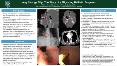
Case Description/Methods: A 36-year-old man presented to the emergency department for multiple gunshot wounds to the head, chest, and extremities. Head CT showed multiple ballistic fragments in the left cerebral hemisphere. Maxillofacial CT showed multiple bilateral facial fractures and scattered ballistic fragments, including one in the nasopharynx. On hospital day ten, the patient had an episode of emesis that was evaluated with an abdominal x-ray that showed a ballistic fragment projecting over the mid thorax along the right lateral margin of the thoracic spine, which was in a different location when compared to previous imaging from presentation. The change in location raised concern that the ballistic fragment in the nasopharynx that was seen on initial maxillofacial imaging had been swallowed and refluxed into the esophagus. Abdominal CT confirmed the location of ballistic fragment in the lower esophagus. An urgent upper endoscopy was performed which showed the bullet fragment at the gastroesophageal junction (Figure). The fragment was pushed into the stomach and removed via Roth net.
Discussion: The timing and necessity of endoscopic intervention for foreign body ingestion depends on multiple factors, including the quality, size, and anatomic location of the object. All ingested objects do not need intervention, but esophageal foreign bodies require urgent intervention within 24 hours of ingestion due to the risk of perforation. The timing of the migration of the ballistic fragment in this patient was not fully understood, and he was unable to aid in spontaneous passage due to an inability to swallow secondary to severe neurologic injury. Therefore, to minimize the risk of perforation, urgent removal of the object was paramount.

Disclosures:
Matthew Houle, MD, John Bloch, MD, MPH, Kaitlin Occhipinti, MD. P0503 - Long Strange Trip: The Story of a Migrating Ballistic Fragment, ACG 2023 Annual Scientific Meeting Abstracts. Vancouver, BC, Canada: American College of Gastroenterology.
Tulane University School of Medicine, New Orleans, LA
Introduction: Foreign body ingestion is a common occurrence, and about 80% of ingestions will pass spontaneously without intervention. However, the remaining approximate 20% of ingestions require an endoscopic procedure. Complications of ingestions include impaction, perforation, or obstruction, and they usually occur at areas of angulation or acute narrowing, such as the gastroesophageal junction. The highest rate of complications occurs in the esophagus, and it is directly related to the dwell time in the esophagus. Thus, urgent evaluation is required for all esophageal foreign bodies. There have been no reported cases of ballistic fragments migrating from the nasopharynx to the esophagus requiring endoscopic removal.
Case Description/Methods: A 36-year-old man presented to the emergency department for multiple gunshot wounds to the head, chest, and extremities. Head CT showed multiple ballistic fragments in the left cerebral hemisphere. Maxillofacial CT showed multiple bilateral facial fractures and scattered ballistic fragments, including one in the nasopharynx. On hospital day ten, the patient had an episode of emesis that was evaluated with an abdominal x-ray that showed a ballistic fragment projecting over the mid thorax along the right lateral margin of the thoracic spine, which was in a different location when compared to previous imaging from presentation. The change in location raised concern that the ballistic fragment in the nasopharynx that was seen on initial maxillofacial imaging had been swallowed and refluxed into the esophagus. Abdominal CT confirmed the location of ballistic fragment in the lower esophagus. An urgent upper endoscopy was performed which showed the bullet fragment at the gastroesophageal junction (Figure). The fragment was pushed into the stomach and removed via Roth net.
Discussion: The timing and necessity of endoscopic intervention for foreign body ingestion depends on multiple factors, including the quality, size, and anatomic location of the object. All ingested objects do not need intervention, but esophageal foreign bodies require urgent intervention within 24 hours of ingestion due to the risk of perforation. The timing of the migration of the ballistic fragment in this patient was not fully understood, and he was unable to aid in spontaneous passage due to an inability to swallow secondary to severe neurologic injury. Therefore, to minimize the risk of perforation, urgent removal of the object was paramount.

Figure: Figure (No 1). Ballistic fragment migration from nasopharynx to esophagus. (A, B) Maxillofacial CT scan demonstrating ballistic fragment in the nasopharynx. (C) Chest radiograph with ballistic fragment projecting over the mid thorax along the right lateral margin of the thoracic spine. (D) Chest CT scan revealing ballistic fragment in lower esophagus. (E) Upper endoscopy showing ballistic wedged at gastroesophageal junction. (F) Upper endoscopy showing ballistic after it was pushed from lower esophagus into the stomach.
Disclosures:
Matthew Houle indicated no relevant financial relationships.
John Bloch indicated no relevant financial relationships.
Kaitlin Occhipinti indicated no relevant financial relationships.
Matthew Houle, MD, John Bloch, MD, MPH, Kaitlin Occhipinti, MD. P0503 - Long Strange Trip: The Story of a Migrating Ballistic Fragment, ACG 2023 Annual Scientific Meeting Abstracts. Vancouver, BC, Canada: American College of Gastroenterology.
