Sunday Poster Session
Category: Esophagus
P0507 - An Unusual Case of Squamous Papilloma Masquerading as Esophageal Malignancy
Sunday, October 22, 2023
3:30 PM - 7:00 PM PT
Location: Exhibit Hall

Has Audio
- WZ
Weidan Zhao, MD
SUNY Downstate Medical Center
Brooklyn, New York
Presenting Author(s)
Weidan Zhao, MD, Evan Grossman, MD
SUNY Downstate Medical Center, Brooklyn, NY
Introduction: Esophageal squamous papilloma (ESP) is a rare small tumor of the esophagus. It is typically asymptomatic, benign, and is found incidentally on upper endoscopy (EGD). We present a case of a large ESP with concomitant candida esophagitis causing severe dysphagia.
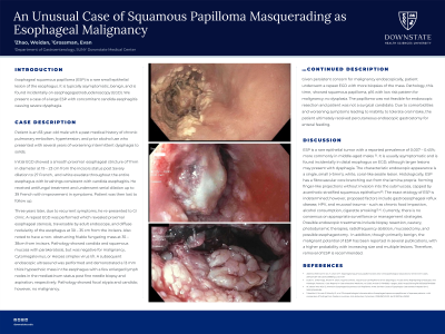
Case Description/Methods: An 83-year-old male with history of pulmonary embolism, hypertension, and prior alcohol use presented with several years of dysphagia to solids. Initial EGD showed an esophageal stricture of 7mm in diameter at 19 – 23cm from the incisors status post Savary dilation to 27 French and white exudate consistent with candida. He received antifungal treatment and underwent serial dilation up to 39 French with improvement in symptoms and was lost to follow up for three years. Due to recurrent symptoms, a repeat EGD was performed which revealed a proximal esophageal stenosis, traversable by adult endoscope, and a non-obstructing friable fungating mass at 35 – 38cm from incisors. Pathology showed candida and squamous mucosa with parakeratosis, but was negative for malignancy, CMV, or HSV I/II. Subsequent endoscopic ultrasound showed a 13mm thick hypoechoic mass in the esophagus with a few enlarged lymph nodes in the mediastinum status post fine needle biopsy and aspiration, respectively. Pathology showed focal atypia and candida. Again, no malignancy. Given endoscopic concern for malignancy, patient underwent a repeat EGD with more biopsies of the mass. Pathology showed squamous papilloma, p16 with low risk pattern for malignancy; no dysplasia. Unable to go through endoscopic or surgical resection, the patient ultimately received percutaneous endoscopic gastrostomy for enteral feeding due to inability to tolerate oral intake.
Discussion: ESP is a rare epithelial tumor with a reported prevalence of 0.007 – 0.45%. It is usually asymptomatic, found incidentally on EGD. The characteristic appearance is a single, < 5mm, white, coral-like sessile lesion. Histologically, ESP has a fibrovascular core capped by acanthotic stratified squamous epithelium. Although of unclear etiology, proposed factors include GERD, HPV, and mucosal trauma. Currently, there is no consensus on appropriate surveillance or management strategies. Possible endoscopic treatments include biopsy resection, cautery, photodynamic therapies, radiofrequency ablation, and mucosectomy. In addition, though primarily benign, the malignant potential of ESP has been reported in several publications, with a higher probability with increasing size and multiple lesions.

Disclosures:
Weidan Zhao, MD, Evan Grossman, MD. P0507 - An Unusual Case of Squamous Papilloma Masquerading as Esophageal Malignancy, ACG 2023 Annual Scientific Meeting Abstracts. Vancouver, BC, Canada: American College of Gastroenterology.
SUNY Downstate Medical Center, Brooklyn, NY
Introduction: Esophageal squamous papilloma (ESP) is a rare small tumor of the esophagus. It is typically asymptomatic, benign, and is found incidentally on upper endoscopy (EGD). We present a case of a large ESP with concomitant candida esophagitis causing severe dysphagia.
Case Description/Methods: An 83-year-old male with history of pulmonary embolism, hypertension, and prior alcohol use presented with several years of dysphagia to solids. Initial EGD showed an esophageal stricture of 7mm in diameter at 19 – 23cm from the incisors status post Savary dilation to 27 French and white exudate consistent with candida. He received antifungal treatment and underwent serial dilation up to 39 French with improvement in symptoms and was lost to follow up for three years. Due to recurrent symptoms, a repeat EGD was performed which revealed a proximal esophageal stenosis, traversable by adult endoscope, and a non-obstructing friable fungating mass at 35 – 38cm from incisors. Pathology showed candida and squamous mucosa with parakeratosis, but was negative for malignancy, CMV, or HSV I/II. Subsequent endoscopic ultrasound showed a 13mm thick hypoechoic mass in the esophagus with a few enlarged lymph nodes in the mediastinum status post fine needle biopsy and aspiration, respectively. Pathology showed focal atypia and candida. Again, no malignancy. Given endoscopic concern for malignancy, patient underwent a repeat EGD with more biopsies of the mass. Pathology showed squamous papilloma, p16 with low risk pattern for malignancy; no dysplasia. Unable to go through endoscopic or surgical resection, the patient ultimately received percutaneous endoscopic gastrostomy for enteral feeding due to inability to tolerate oral intake.
Discussion: ESP is a rare epithelial tumor with a reported prevalence of 0.007 – 0.45%. It is usually asymptomatic, found incidentally on EGD. The characteristic appearance is a single, < 5mm, white, coral-like sessile lesion. Histologically, ESP has a fibrovascular core capped by acanthotic stratified squamous epithelium. Although of unclear etiology, proposed factors include GERD, HPV, and mucosal trauma. Currently, there is no consensus on appropriate surveillance or management strategies. Possible endoscopic treatments include biopsy resection, cautery, photodynamic therapies, radiofrequency ablation, and mucosectomy. In addition, though primarily benign, the malignant potential of ESP has been reported in several publications, with a higher probability with increasing size and multiple lesions.

Figure: Endoscopic image of esophageal squamous papilloma
Disclosures:
Weidan Zhao indicated no relevant financial relationships.
Evan Grossman indicated no relevant financial relationships.
Weidan Zhao, MD, Evan Grossman, MD. P0507 - An Unusual Case of Squamous Papilloma Masquerading as Esophageal Malignancy, ACG 2023 Annual Scientific Meeting Abstracts. Vancouver, BC, Canada: American College of Gastroenterology.
