Sunday Poster Session
Category: Esophagus
P0508 - Uncommon Cause of Esophageal Pneumatosis With Endoscopic Ultrasound Findings
Sunday, October 22, 2023
3:30 PM - 7:00 PM PT
Location: Exhibit Hall

Has Audio

Kwabena O. Asafo-Agyei, MD
Christus Highland Hospital
Shreveport, LA
Presenting Author(s)
Kwabena O. Asafo-Agyei, MD1, Raymond Kim, MD, DM2
1Christus Highland Hospital, Shreveport, LA; 2University of Maryland Medical Center, Baltimore, MD
Introduction: Pneumatosis intestinalis is the presence of gas in the bowel wall and can occur anywhere in the bowel; the esophagus, stomach, small intestines, and colon. Isolated esophageal pneumatosis is rare, but none reports the Endoscopic Ultrasound (EUS) imaging findings of this occurrence.
Case Description/Methods: A 51-year-old man presented to the clinic with intractable hiccups for 2 years after coronary artery bypass graft surgery (CABG) in 2014. His hiccups started immediately after CABG. Hiccups were associated with most of his meals and were relieved by inducing self-induced vomiting by self-gagging. The patient felt bloated but denied any other symptoms. He had initially been medicated on chlorpromazine and metoclopramide but had no improvement. His previous abdominal and chest non-contrast computed imaging were unrevealing for the cause of the hiccups. In addition, the patient had and normal gastric emptying study.
He had Esophagogastroduodenoscopy (EGD), which revealed subepithelial nodules in the mid-esophagus (Figure 1). The appearance of the esophagus suggested pneumatosis of the esophageal wall and was hence not biopsied. Findings during EGD revealed erythematous mucosa in the gastric body, antrum, and first part of the duodenum, which was then biopsied. The biopsy report showed normal mucosa and negative Helicobacter pylori staining.
Follow-up EGD with Endoscopic Ultrasound (EUS) a week later revealed blebs in the esophagus consistent with pneumatosis (Figure 2). An intramural (subepithelial) lesion in the esophagus appeared to originate within the submucosa (layer 3). The patient had pneumatosis esophagitis with mild gastritis of the antrum and otherwise normal EGD findings.
Discussion: There have been 2 postulated hypotheses of pathogenesis. This includes the mechanical and bacteria theory. Lederman reported 13% of patients undergoing CABG developed new peripheral nervous system complications postoperatively. We hypothesize that this could be the etiology of his intractable hiccups. Also, hiccups are associated with increased intrathoracic pressure and could lead to esophageal pneumatosis, as postulated by the mechanical theory, from increased intraluminal pressure and possible mucosal disruption.
Endoscopic Ultrasound findings of esophageal pneumatosis have never been published, and sensitivities and specificities of recognition of these findings are unknown. However, identifying findings is critical in avoiding biopsy and its attended complications of perforation and mediastinitis.

Disclosures:
Kwabena O. Asafo-Agyei, MD1, Raymond Kim, MD, DM2. P0508 - Uncommon Cause of Esophageal Pneumatosis With Endoscopic Ultrasound Findings, ACG 2023 Annual Scientific Meeting Abstracts. Vancouver, BC, Canada: American College of Gastroenterology.
1Christus Highland Hospital, Shreveport, LA; 2University of Maryland Medical Center, Baltimore, MD
Introduction: Pneumatosis intestinalis is the presence of gas in the bowel wall and can occur anywhere in the bowel; the esophagus, stomach, small intestines, and colon. Isolated esophageal pneumatosis is rare, but none reports the Endoscopic Ultrasound (EUS) imaging findings of this occurrence.
Case Description/Methods: A 51-year-old man presented to the clinic with intractable hiccups for 2 years after coronary artery bypass graft surgery (CABG) in 2014. His hiccups started immediately after CABG. Hiccups were associated with most of his meals and were relieved by inducing self-induced vomiting by self-gagging. The patient felt bloated but denied any other symptoms. He had initially been medicated on chlorpromazine and metoclopramide but had no improvement. His previous abdominal and chest non-contrast computed imaging were unrevealing for the cause of the hiccups. In addition, the patient had and normal gastric emptying study.
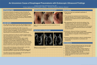
He had Esophagogastroduodenoscopy (EGD), which revealed subepithelial nodules in the mid-esophagus (Figure 1). The appearance of the esophagus suggested pneumatosis of the esophageal wall and was hence not biopsied. Findings during EGD revealed erythematous mucosa in the gastric body, antrum, and first part of the duodenum, which was then biopsied. The biopsy report showed normal mucosa and negative Helicobacter pylori staining.
Follow-up EGD with Endoscopic Ultrasound (EUS) a week later revealed blebs in the esophagus consistent with pneumatosis (Figure 2). An intramural (subepithelial) lesion in the esophagus appeared to originate within the submucosa (layer 3). The patient had pneumatosis esophagitis with mild gastritis of the antrum and otherwise normal EGD findings.
Discussion: There have been 2 postulated hypotheses of pathogenesis. This includes the mechanical and bacteria theory. Lederman reported 13% of patients undergoing CABG developed new peripheral nervous system complications postoperatively. We hypothesize that this could be the etiology of his intractable hiccups. Also, hiccups are associated with increased intrathoracic pressure and could lead to esophageal pneumatosis, as postulated by the mechanical theory, from increased intraluminal pressure and possible mucosal disruption.
Endoscopic Ultrasound findings of esophageal pneumatosis have never been published, and sensitivities and specificities of recognition of these findings are unknown. However, identifying findings is critical in avoiding biopsy and its attended complications of perforation and mediastinitis.

Figure: Figure 1 shows subepithelial nodules in the mid-esophagus
Figure 2 revealed blebs in the esophagus consistent with pneumatosis and an intramural (subepithelial) lesion in the esophagus appeared to originate within the submucosa (layer 3).
Figure 2 revealed blebs in the esophagus consistent with pneumatosis and an intramural (subepithelial) lesion in the esophagus appeared to originate within the submucosa (layer 3).
Disclosures:
Kwabena Asafo-Agyei indicated no relevant financial relationships.
Raymond Kim: Apollo Endosurgery – Consultant. Cook medical – Consultant.
Kwabena O. Asafo-Agyei, MD1, Raymond Kim, MD, DM2. P0508 - Uncommon Cause of Esophageal Pneumatosis With Endoscopic Ultrasound Findings, ACG 2023 Annual Scientific Meeting Abstracts. Vancouver, BC, Canada: American College of Gastroenterology.
