Sunday Poster Session
Category: Esophagus
P0516 - Disappearing Dysphagia and the Spontaneous Intra-Esophageal Hematoma
Sunday, October 22, 2023
3:30 PM - 7:00 PM PT
Location: Exhibit Hall

Has Audio

Austin Wayne Wheeler, MD
LSU Health Sciences Center
New Orleans, LA
Presenting Author(s)
Award: Presidential Poster Award
Austin Wayne Wheeler, MD1, Stephen Wayne. Landreneau, MD2
1LSU Health Sciences Center, New Orleans, LA; 2Louisiana State University, New Orleans, LA
Introduction: The pathophysiology of dysphagia can involve mechanical, structural, or infectious etiologies. This case represents a rare cause of dysphagia due to a spontaneous intra-esophageal hematoma.
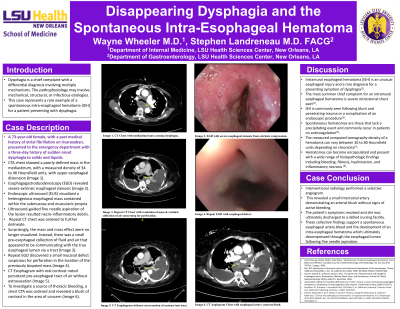
Case Description/Methods: A 73-year-old female with a history of atrial fibrillation, on rivaroxaban, presented with a three-day history of acute dysphagia to solids and liquids. Chest CT angiography showed an esophageal mass with a density of 34-46 Hounsfield units. EGD revealed severe extrinsic esophageal stenosis. Endoscopic ultrasound visualized a heterogenous mass within the submucosa and muscularis propria. Ultrasound-guided fine needle aspiration of the mass resulted necro-inflammatory debris. Repeat CT chest was ordered to further delineate the findings. Surprisingly, the mass had disappeared. Instead, a pre-esophageal lumen possibly communicating with the true esophageal lumen via a tract was seen. Repeat EGD discovered a small mucosal defect in the location of the previous biopsy. CT esophagram was performed without extravasation of contrast into the false lumen. To investigate a source of thoracic bleeding, a chest CTA was ordered and revealed a blush of contrast from a mid-esophageal artery. Interventional radiology performed a selective catheterization that showed a late blush of contrast in the area of concern, corresponding with the CT findings. No active bleeding was identified. The patient’s dysphagia improved, and she was tolerating a regular diet prior to discharge. These findings support a spontaneous esophageal artery bleed and the development of an intra-esophageal hematoma which ultimately decompressed through the esophageal lumen without further intervention.
Discussion: Intramural esophageal hematoma (IEH) is an unusual esophageal injury and a rare diagnosis for dysphagia. The most common chief complaint for IEH is severe retrosternal chest pain. IEH is commonly seen following blunt and penetrating trauma or as a complication of an endoscopic procedure. Spontaneous hematomas are those that lack a precipitating event and can occur in patients on anticoagulation. The measured CT density of a hematoma is between 30 to 80 Hounsfield units depending on chronicity. Hematomas can become encapsulated and present with a range of histopathologic findings including bleeding, fibrosis, hyalinization, and inflammatory necrosis. This report is a unique case of a patient on anticoagulation presenting with a spontaneous IEH in the absence of trauma or prior endoscopic procedure.

Disclosures:
Austin Wayne Wheeler, MD1, Stephen Wayne. Landreneau, MD2. P0516 - Disappearing Dysphagia and the Spontaneous Intra-Esophageal Hematoma, ACG 2023 Annual Scientific Meeting Abstracts. Vancouver, BC, Canada: American College of Gastroenterology.
Austin Wayne Wheeler, MD1, Stephen Wayne. Landreneau, MD2
1LSU Health Sciences Center, New Orleans, LA; 2Louisiana State University, New Orleans, LA
Introduction: The pathophysiology of dysphagia can involve mechanical, structural, or infectious etiologies. This case represents a rare cause of dysphagia due to a spontaneous intra-esophageal hematoma.
Case Description/Methods: A 73-year-old female with a history of atrial fibrillation, on rivaroxaban, presented with a three-day history of acute dysphagia to solids and liquids. Chest CT angiography showed an esophageal mass with a density of 34-46 Hounsfield units. EGD revealed severe extrinsic esophageal stenosis. Endoscopic ultrasound visualized a heterogenous mass within the submucosa and muscularis propria. Ultrasound-guided fine needle aspiration of the mass resulted necro-inflammatory debris. Repeat CT chest was ordered to further delineate the findings. Surprisingly, the mass had disappeared. Instead, a pre-esophageal lumen possibly communicating with the true esophageal lumen via a tract was seen. Repeat EGD discovered a small mucosal defect in the location of the previous biopsy. CT esophagram was performed without extravasation of contrast into the false lumen. To investigate a source of thoracic bleeding, a chest CTA was ordered and revealed a blush of contrast from a mid-esophageal artery. Interventional radiology performed a selective catheterization that showed a late blush of contrast in the area of concern, corresponding with the CT findings. No active bleeding was identified. The patient’s dysphagia improved, and she was tolerating a regular diet prior to discharge. These findings support a spontaneous esophageal artery bleed and the development of an intra-esophageal hematoma which ultimately decompressed through the esophageal lumen without further intervention.
Discussion: Intramural esophageal hematoma (IEH) is an unusual esophageal injury and a rare diagnosis for dysphagia. The most common chief complaint for IEH is severe retrosternal chest pain. IEH is commonly seen following blunt and penetrating trauma or as a complication of an endoscopic procedure. Spontaneous hematomas are those that lack a precipitating event and can occur in patients on anticoagulation. The measured CT density of a hematoma is between 30 to 80 Hounsfield units depending on chronicity. Hematomas can become encapsulated and present with a range of histopathologic findings including bleeding, fibrosis, hyalinization, and inflammatory necrosis. This report is a unique case of a patient on anticoagulation presenting with a spontaneous IEH in the absence of trauma or prior endoscopic procedure.

Figure: Figure A: CTA Chest with mediastinal mass causing dysphagia.
Figure B: EGD revealing severe extrinsic esophageal stenosis.
Figure C: EUS showing esophageal mass within submucosa and muscularis propria.
Figure D: Repeat CT chest with resolution of mass and residual collection of air.
Figure E: Repeat EGD with mucosal defect at site of biopsy.
Figure F: CT esophagram without extravasation of contrast into false lumen.
Figure G: CTA chest with esophageal artery contrast blush.
Figure H: Arterial blush seen on aortogram with selective catheterization of intercostal artery.
Disclosures:
Austin Wayne Wheeler indicated no relevant financial relationships.
Stephen Landreneau indicated no relevant financial relationships.
Austin Wayne Wheeler, MD1, Stephen Wayne. Landreneau, MD2. P0516 - Disappearing Dysphagia and the Spontaneous Intra-Esophageal Hematoma, ACG 2023 Annual Scientific Meeting Abstracts. Vancouver, BC, Canada: American College of Gastroenterology.

