Sunday Poster Session
Category: Esophagus
P0519 - Black Esophagus: An Unusual Accomplice to Acute Esophageal Necrosis
Sunday, October 22, 2023
3:30 PM - 7:00 PM PT
Location: Exhibit Hall

Has Audio
- SR
Shanjeev Kumar Rajeshkumar Chitra, MD
Mercy Catholic Medical Center
Lansdowne, PA
Presenting Author(s)
Shanjeev Kumar Rajeshkumar Chitra, MD, Nithya Ramesh, MD, Sherley Chhibber, MD, Sruthi Sekaran, MD, Mansi Naik, MD, Tharika Shraddha, MD
Mercy Catholic Medical Center, Darby, PA
Introduction: Circumferential black discoloration of the esophageal mucosa is concerning for acute esophageal necrosis (AEN), a rare condition resulting from ischemic insult to the mucosa from hypoperfusion or thromboembolic events. Diabetic ketoacidosis (DKA) being a low perfusion state is known to predispose to AEN. But here we describe an interesting case of AEN in a young man with DKA, with histopathology revealing an unusual accomplice.
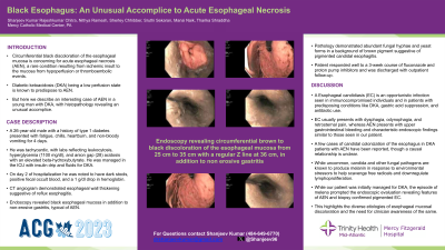
Case Description/Methods: A 26-year-old male with a history of type 1 diabetes presented with fatigue, chills, heartburn, and non-bloody vomiting for 4 days. He was tachycardic, with labs reflecting leukocytosis, hyperglycemia (1100 mg/dl), and anion gap (28) acidosis with an elevated beta-hydroxybutyrate. He was managed in the ICU with insulin drip and fluids for DKA. On day 2 of hospitalization he was noted to have dark stools, positive fecal occult blood, and a 1 g/dl drop in hemoglobin. CT angiogram demonstrated esophageal wall thickening suggestive of reflux esophagitis. Endoscopy revealed black colored esophageal mucosa in addition to non-erosive gastritis, typical of AEN. Pathology demonstrated abundant fungal hyphae and yeast forms in a background of brown pigment suggestive of pigmented candidal esophagitis. Patient responded well to a 3-week course of fluconazole and proton pump inhibitors and was discharged with outpatient follow-up.
Discussion: Esophageal candidiasis (EC) is an opportunistic infection seen in immunocompromised individuals and in patients with predisposing conditions like DKA, gastric acid suppression, and antibiotic use. EC usually presents with dysphagia, odynophagia, and retrosternal pain, whereas AEN presents with upper gastrointestinal bleeding and characteristic endoscopic findings similar to those seen in our patient. A few cases of candidal colonization of the esophagus in DKA patients with AEN have been reported, though a causal relationship is unclear. While uncommon, candida and other fungal pathogens are known to produce melanin in response to environmental stressors to help scavenge free radicals and downregulate lymphoproliferation. While our patient was initially managed for DKA, the episode of melena prompted the endoscopic evaluation revealing features of AEN and biopsy confirmed pigmented EC. This highlights the diverse etiologies of esophageal mucosal discoloration and the need for clinician awareness of the same.

Disclosures:
Shanjeev Kumar Rajeshkumar Chitra, MD, Nithya Ramesh, MD, Sherley Chhibber, MD, Sruthi Sekaran, MD, Mansi Naik, MD, Tharika Shraddha, MD. P0519 - Black Esophagus: An Unusual Accomplice to Acute Esophageal Necrosis, ACG 2023 Annual Scientific Meeting Abstracts. Vancouver, BC, Canada: American College of Gastroenterology.
Mercy Catholic Medical Center, Darby, PA
Introduction: Circumferential black discoloration of the esophageal mucosa is concerning for acute esophageal necrosis (AEN), a rare condition resulting from ischemic insult to the mucosa from hypoperfusion or thromboembolic events. Diabetic ketoacidosis (DKA) being a low perfusion state is known to predispose to AEN. But here we describe an interesting case of AEN in a young man with DKA, with histopathology revealing an unusual accomplice.
Case Description/Methods: A 26-year-old male with a history of type 1 diabetes presented with fatigue, chills, heartburn, and non-bloody vomiting for 4 days. He was tachycardic, with labs reflecting leukocytosis, hyperglycemia (1100 mg/dl), and anion gap (28) acidosis with an elevated beta-hydroxybutyrate. He was managed in the ICU with insulin drip and fluids for DKA. On day 2 of hospitalization he was noted to have dark stools, positive fecal occult blood, and a 1 g/dl drop in hemoglobin. CT angiogram demonstrated esophageal wall thickening suggestive of reflux esophagitis. Endoscopy revealed black colored esophageal mucosa in addition to non-erosive gastritis, typical of AEN. Pathology demonstrated abundant fungal hyphae and yeast forms in a background of brown pigment suggestive of pigmented candidal esophagitis. Patient responded well to a 3-week course of fluconazole and proton pump inhibitors and was discharged with outpatient follow-up.
Discussion: Esophageal candidiasis (EC) is an opportunistic infection seen in immunocompromised individuals and in patients with predisposing conditions like DKA, gastric acid suppression, and antibiotic use. EC usually presents with dysphagia, odynophagia, and retrosternal pain, whereas AEN presents with upper gastrointestinal bleeding and characteristic endoscopic findings similar to those seen in our patient. A few cases of candidal colonization of the esophagus in DKA patients with AEN have been reported, though a causal relationship is unclear. While uncommon, candida and other fungal pathogens are known to produce melanin in response to environmental stressors to help scavenge free radicals and downregulate lymphoproliferation. While our patient was initially managed for DKA, the episode of melena prompted the endoscopic evaluation revealing features of AEN and biopsy confirmed pigmented EC. This highlights the diverse etiologies of esophageal mucosal discoloration and the need for clinician awareness of the same.

Figure: Endoscopy revealing circumferential black discoloration of the esophageal mucosa from 25 cm to 35 cm with a regular Z line at 36 cm, in addition to non-erosive gastritis
Disclosures:
Shanjeev Kumar Rajeshkumar Chitra indicated no relevant financial relationships.
Nithya Ramesh indicated no relevant financial relationships.
Sherley Chhibber indicated no relevant financial relationships.
Sruthi Sekaran indicated no relevant financial relationships.
Mansi Naik indicated no relevant financial relationships.
Tharika Shraddha indicated no relevant financial relationships.
Shanjeev Kumar Rajeshkumar Chitra, MD, Nithya Ramesh, MD, Sherley Chhibber, MD, Sruthi Sekaran, MD, Mansi Naik, MD, Tharika Shraddha, MD. P0519 - Black Esophagus: An Unusual Accomplice to Acute Esophageal Necrosis, ACG 2023 Annual Scientific Meeting Abstracts. Vancouver, BC, Canada: American College of Gastroenterology.
