Sunday Poster Session
Category: General Endoscopy
P0579 - A Colorful Case of Esophageal Blebs
Sunday, October 22, 2023
3:30 PM - 7:00 PM PT
Location: Exhibit Hall

Has Audio

Katelyn Madigan, MD
Atrium Health Wake Forest Baptist
Winston-Salem, NC
Presenting Author(s)
Katelyn Madigan, MD, Ann Honor, MD
Atrium Health Wake Forest Baptist, Winston-Salem, NC
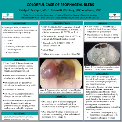
Introduction: Esophageal blebs, also known as submucosal esophageal hematomas, are rare endoscopic findings. Proposed etiologies for this uncommon phenomenon include injury secondary to trauma, toxins, or following endoscopic interventions. Case reports conjecture that thrombocytopenia or copper excess can contribute to this finding. Here we present a case of a 59-year-old female with decompensated Wilson’s cirrhosis (ascites, hepatic encephalopathy, and non-bleeding esophageal varices) who presented for evaluation of subacute dysphagia to solids and liquids.
Case Description/Methods: On presentation, the patient was afebrile and hemodynamically stable. She was maintained on a stable dose of trientine, and denied NSAID use, recent medication changes, or intercurrent illnesses. Her physical exam had prominent ascites, lower extremity edema, peripheral muscular atrophy, diffuse ecchymoses, and spider angiomas. Labs noted sodium 126, BUN 29, creatinine 1.0, tBili 6.7, dBili 3.1, albumin 2.7, ALP 88, AST 20, ALT 16, ammonia 38, INR 1.7, hemoglobin 8.8, MCV 110, platelets 25k (confirmed on repeat), haptoglobin 30, LDH 153. MELD-Na 27. A 24-hour urine copper was elevated at 356 ug/24h. A non-contrast CT scan (Image 1) revealed cirrhotic liver morphology and prominent splenomegaly - felt to be the etiology of her severe thrombocytopenia. An upper endoscopy in 2020 revealed grade 1-2 distal esophageal varices partially collapsible upon insufflation and minimal portal gastropathy. A repeat upper endoscopy was obtained to evaluate her dysphagia, which revealed new findings of extensive and numerous non-bleeding yellow and dark red esophageal blebs (Image 2).
Discussion: Risk factors for esophageal blebs include female gender, coagulopathy, and increased intra-esophageal pressure, such as induced by vomiting. Case reports (Sharma et al. Radiol Case Rep 12:278-280, 2017; Ashman et al. Gastrointest Radiol 3:115–118, 1978) have proposed that thrombocytopenia leading to intramural hemorrhage is a factor that contributes to hemorrhagic bleb formation. Relevant to our patient, elevated copper levels may induce submucosal inflammation and fibrosis (Sachdev et al. Int J Dent 2018, Jun 26:3472087), which could have compromised the integrity of her esophageal submucosa and contributed to the formation of the yellow, presumably serous, blebs. Management of submucosal esophageal hematomas aims to treat the underlying causes. Correction of coagulopathy, such as thrombocytopenia, or copper overload should be attempted for treatment.

Disclosures:
Katelyn Madigan, MD, Ann Honor, MD. P0579 - A Colorful Case of Esophageal Blebs, ACG 2023 Annual Scientific Meeting Abstracts. Vancouver, BC, Canada: American College of Gastroenterology.
Atrium Health Wake Forest Baptist, Winston-Salem, NC
Introduction: Esophageal blebs, also known as submucosal esophageal hematomas, are rare endoscopic findings. Proposed etiologies for this uncommon phenomenon include injury secondary to trauma, toxins, or following endoscopic interventions. Case reports conjecture that thrombocytopenia or copper excess can contribute to this finding. Here we present a case of a 59-year-old female with decompensated Wilson’s cirrhosis (ascites, hepatic encephalopathy, and non-bleeding esophageal varices) who presented for evaluation of subacute dysphagia to solids and liquids.
Case Description/Methods: On presentation, the patient was afebrile and hemodynamically stable. She was maintained on a stable dose of trientine, and denied NSAID use, recent medication changes, or intercurrent illnesses. Her physical exam had prominent ascites, lower extremity edema, peripheral muscular atrophy, diffuse ecchymoses, and spider angiomas. Labs noted sodium 126, BUN 29, creatinine 1.0, tBili 6.7, dBili 3.1, albumin 2.7, ALP 88, AST 20, ALT 16, ammonia 38, INR 1.7, hemoglobin 8.8, MCV 110, platelets 25k (confirmed on repeat), haptoglobin 30, LDH 153. MELD-Na 27. A 24-hour urine copper was elevated at 356 ug/24h. A non-contrast CT scan (Image 1) revealed cirrhotic liver morphology and prominent splenomegaly - felt to be the etiology of her severe thrombocytopenia. An upper endoscopy in 2020 revealed grade 1-2 distal esophageal varices partially collapsible upon insufflation and minimal portal gastropathy. A repeat upper endoscopy was obtained to evaluate her dysphagia, which revealed new findings of extensive and numerous non-bleeding yellow and dark red esophageal blebs (Image 2).
Discussion: Risk factors for esophageal blebs include female gender, coagulopathy, and increased intra-esophageal pressure, such as induced by vomiting. Case reports (Sharma et al. Radiol Case Rep 12:278-280, 2017; Ashman et al. Gastrointest Radiol 3:115–118, 1978) have proposed that thrombocytopenia leading to intramural hemorrhage is a factor that contributes to hemorrhagic bleb formation. Relevant to our patient, elevated copper levels may induce submucosal inflammation and fibrosis (Sachdev et al. Int J Dent 2018, Jun 26:3472087), which could have compromised the integrity of her esophageal submucosa and contributed to the formation of the yellow, presumably serous, blebs. Management of submucosal esophageal hematomas aims to treat the underlying causes. Correction of coagulopathy, such as thrombocytopenia, or copper overload should be attempted for treatment.

Figure: Image 1: Numerous yellow and dark red non-bleeding esophageal blebs
Disclosures:
Katelyn Madigan indicated no relevant financial relationships.
Ann Honor indicated no relevant financial relationships.
Katelyn Madigan, MD, Ann Honor, MD. P0579 - A Colorful Case of Esophageal Blebs, ACG 2023 Annual Scientific Meeting Abstracts. Vancouver, BC, Canada: American College of Gastroenterology.
