Sunday Poster Session
Category: General Endoscopy
P0588 - Primary Systemic AL Amyloidosis Diagnosed by Gastric Ulcer Biopsy: A Case Report
Sunday, October 22, 2023
3:30 PM - 7:00 PM PT
Location: Exhibit Hall

Has Audio

Manasa Narasimman, MD
University of Texas Southwestern Medical Center Dallas
Dallas, TX
Presenting Author(s)
Manasa Narasimman, MD, Sakina Neemuchwala, BS, Rozina Mithani, BA, MD
University of Texas Southwestern Medical Center Dallas, Dallas, TX
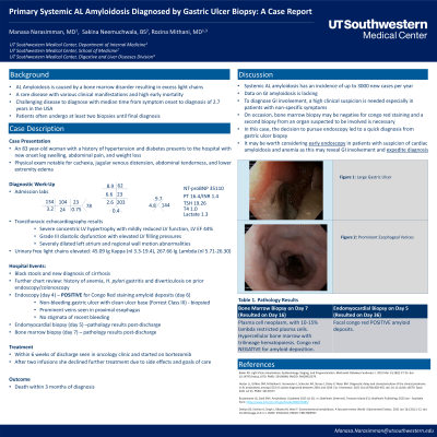
Introduction: AL amyloidosis is a rare and challenging disease to diagnose with diverse clinical manifestations. The median time from symptom onset to diagnosis in the USA is 2.7 years and many undergo at least 2 biopsies prior to diagnosis. We present a case of systemic AL amyloidosis diagnosed by endoscopy soon after initial presentation.
Case Description/Methods: An 83 y/o F with hypertension and diabetes presented to the hospital with new onset leg swelling, abdominal pain, and weight loss. Initial exam was notable for cachexia, jugular venous distension, abdominal tenderness, and lower extremity edema. Labs notable for NT-proBNP 35110, Hgb 9.7, AST 62, ALP 203, and INR 1.4. Transthoracic echocardiography revealed severe concentric left ventricle hypertrophy, an ejection fraction of 44%, and grade III diastolic dysfunction with elevated left ventricle filling pressures. Initial differential included new onset heart failure due to uncontrolled hypertension vs an infiltrative process. Free light chains, SPEP, and UPEP were pending. Prior endoscopy for anemia in the year prior showed H. pylori gastritis, a 4mm polyp, diverticulosis, and hemorrhoids. While admitted she had black stools and a new diagnosis of cirrhosis. Endoscopy was pursued on day 4 of admission and showed a large clean-based pre-pyloric ulcer. She was started on treatment for H. pylori while awaiting biopsy results. Her free light chains returned with an elevated kappa to lambda ratio concerning for AL amyloidosis. She underwent endomyocardial biopsy on day 5 and her gastric ulcer pathology returned on day 6 with Congo red stain positive amyloid deposits. She then underwent bone marrow biopsy after which she was discharged to follow up outpatient. Within the next 6 weeks she followed up with oncology and was started on bortezomib despite not having cardiac biopsy results. Subsequently her cardiac biopsy revealed Congo red positive amyloid deposits while her bone marrow was Congo red negative. Due to treatment side effects the patient chose to not pursue chemotherapy after two infusions, and she passed within 3 months of her diagnosis.
Discussion: This case demonstrates the use of endoscopy for an early diagnosis of amyloidosis, despite the lack of cardiac biopsy. While being unconventional, this case illustrates how why it may be worth considering early endoscopy in patients with suspicion of cardiac amyloidosis and anemia as this may reveal GI involvement and expedite diagnosis and subsequent treatment options.

Disclosures:
Manasa Narasimman, MD, Sakina Neemuchwala, BS, Rozina Mithani, BA, MD. P0588 - Primary Systemic AL Amyloidosis Diagnosed by Gastric Ulcer Biopsy: A Case Report, ACG 2023 Annual Scientific Meeting Abstracts. Vancouver, BC, Canada: American College of Gastroenterology.
University of Texas Southwestern Medical Center Dallas, Dallas, TX
Introduction: AL amyloidosis is a rare and challenging disease to diagnose with diverse clinical manifestations. The median time from symptom onset to diagnosis in the USA is 2.7 years and many undergo at least 2 biopsies prior to diagnosis. We present a case of systemic AL amyloidosis diagnosed by endoscopy soon after initial presentation.
Case Description/Methods: An 83 y/o F with hypertension and diabetes presented to the hospital with new onset leg swelling, abdominal pain, and weight loss. Initial exam was notable for cachexia, jugular venous distension, abdominal tenderness, and lower extremity edema. Labs notable for NT-proBNP 35110, Hgb 9.7, AST 62, ALP 203, and INR 1.4. Transthoracic echocardiography revealed severe concentric left ventricle hypertrophy, an ejection fraction of 44%, and grade III diastolic dysfunction with elevated left ventricle filling pressures. Initial differential included new onset heart failure due to uncontrolled hypertension vs an infiltrative process. Free light chains, SPEP, and UPEP were pending. Prior endoscopy for anemia in the year prior showed H. pylori gastritis, a 4mm polyp, diverticulosis, and hemorrhoids. While admitted she had black stools and a new diagnosis of cirrhosis. Endoscopy was pursued on day 4 of admission and showed a large clean-based pre-pyloric ulcer. She was started on treatment for H. pylori while awaiting biopsy results. Her free light chains returned with an elevated kappa to lambda ratio concerning for AL amyloidosis. She underwent endomyocardial biopsy on day 5 and her gastric ulcer pathology returned on day 6 with Congo red stain positive amyloid deposits. She then underwent bone marrow biopsy after which she was discharged to follow up outpatient. Within the next 6 weeks she followed up with oncology and was started on bortezomib despite not having cardiac biopsy results. Subsequently her cardiac biopsy revealed Congo red positive amyloid deposits while her bone marrow was Congo red negative. Due to treatment side effects the patient chose to not pursue chemotherapy after two infusions, and she passed within 3 months of her diagnosis.
Discussion: This case demonstrates the use of endoscopy for an early diagnosis of amyloidosis, despite the lack of cardiac biopsy. While being unconventional, this case illustrates how why it may be worth considering early endoscopy in patients with suspicion of cardiac amyloidosis and anemia as this may reveal GI involvement and expedite diagnosis and subsequent treatment options.

Figure: Gastric Ulcer with Amyloidosis
Disclosures:
Manasa Narasimman indicated no relevant financial relationships.
Sakina Neemuchwala indicated no relevant financial relationships.
Rozina Mithani indicated no relevant financial relationships.
Manasa Narasimman, MD, Sakina Neemuchwala, BS, Rozina Mithani, BA, MD. P0588 - Primary Systemic AL Amyloidosis Diagnosed by Gastric Ulcer Biopsy: A Case Report, ACG 2023 Annual Scientific Meeting Abstracts. Vancouver, BC, Canada: American College of Gastroenterology.
