Sunday Poster Session
Category: GI Bleeding
P0619 - Recurrent Renal Cell Carcinoma and Hematochezia: An Extraordinary Presentation
Sunday, October 22, 2023
3:30 PM - 7:00 PM PT
Location: Exhibit Hall

Has Audio

Thomas Smith, MD
Mayo Clinic School of Graduate Medical Education
Rochester, Minnesota
Presenting Author(s)
Stephanie Hansel, MD, MS1, David Chiang, MD, PhD1, Thomas Smith, MD2
1Mayo Clinic, Rochester, MN; 2Mayo Clinic School of Graduate Medical Education, Rochester, MN
Introduction: Renal cell carcinoma (RCC) is the most common malignancy of the kidney, with metastasis present at diagnosis in up to 30% of cases. Even after nephrectomy, the rate of metastatic recurrence is nearly 40% with one in ten developing late metastatic disease after 5 years. Common metastasis sites are lung (50-60%), bone (30-40%), liver (30-40%) and brain (5%). Clinically apparent colon metastasis is exceedingly rare, with a comprehensive literature review in 2021 yielding only 26 cases. Colon metastasis is typically associated with disseminated metastatic disease.
Case Description/Methods: We present the case of a 68-year-old white male with new onset hematochezia and anemia. He presented to the ED with two days of grossly bloody stools and fatigue. Relevant medical comorbidities include prior renal cell carcinoma with left radical nephrectomy performed in 2017, transurethral resection of the prostate performed in 2019, Roux-en-Y bypass, and current Xarelto use following a pulmonary embolism in 2021. Upon presentation, he was found to be hypotensive with new anemia (HGB 9.1 g/dL). Following crystalloid resuscitation, he remained hemodynamically stable. Physical exam including abdominal palpation and auscultation was unremarkable. CT of the abdomen demonstrated a 4x3x3.2 cm enhancing sigmoid mass and liver hypodensities concerning for metastasis. Xarelto was discontinued, and the patient was admitted. Colonoscopy demonstrated polypoid mass with ulcerated and friable elements in the sigmoid colon. Biopsies revealed recurrent renal cell carcinoma. Based on likely metastasis in the liver and lung, resection was not offered by colorectal surgery. The inpatient GI team did not feel further endoscopic intervention would provide benefit. Interventional radiology was unable to localize bleeding vessels, and arterial embolization was not pursued. The patient underwent palliative radiation to decrease further bleeding risk. Medical oncology recommended chemotherapy with Nivolumab, worried that a typical two-agent regimen may worsen bleeding. During treatment, he developed persistent fatigue, weakness and falls in his group home. After 4 treatments over 8 weeks, CT showed growth of both mass and metastases. After 9 weeks, the patient’s hematochezia and hypotension returned, and the decision was made to transition to comfort care.
Discussion: This case highlights an uncommon presentation of RCC recurrence, as well as important treatment considerations in the presence of clinically significant colonic bleeding.

Disclosures:
Stephanie Hansel, MD, MS1, David Chiang, MD, PhD1, Thomas Smith, MD2. P0619 - Recurrent Renal Cell Carcinoma and Hematochezia: An Extraordinary Presentation, ACG 2023 Annual Scientific Meeting Abstracts. Vancouver, BC, Canada: American College of Gastroenterology.
1Mayo Clinic, Rochester, MN; 2Mayo Clinic School of Graduate Medical Education, Rochester, MN
Introduction: Renal cell carcinoma (RCC) is the most common malignancy of the kidney, with metastasis present at diagnosis in up to 30% of cases. Even after nephrectomy, the rate of metastatic recurrence is nearly 40% with one in ten developing late metastatic disease after 5 years. Common metastasis sites are lung (50-60%), bone (30-40%), liver (30-40%) and brain (5%). Clinically apparent colon metastasis is exceedingly rare, with a comprehensive literature review in 2021 yielding only 26 cases. Colon metastasis is typically associated with disseminated metastatic disease.
Case Description/Methods: We present the case of a 68-year-old white male with new onset hematochezia and anemia. He presented to the ED with two days of grossly bloody stools and fatigue. Relevant medical comorbidities include prior renal cell carcinoma with left radical nephrectomy performed in 2017, transurethral resection of the prostate performed in 2019, Roux-en-Y bypass, and current Xarelto use following a pulmonary embolism in 2021. Upon presentation, he was found to be hypotensive with new anemia (HGB 9.1 g/dL). Following crystalloid resuscitation, he remained hemodynamically stable. Physical exam including abdominal palpation and auscultation was unremarkable. CT of the abdomen demonstrated a 4x3x3.2 cm enhancing sigmoid mass and liver hypodensities concerning for metastasis. Xarelto was discontinued, and the patient was admitted. Colonoscopy demonstrated polypoid mass with ulcerated and friable elements in the sigmoid colon. Biopsies revealed recurrent renal cell carcinoma. Based on likely metastasis in the liver and lung, resection was not offered by colorectal surgery. The inpatient GI team did not feel further endoscopic intervention would provide benefit. Interventional radiology was unable to localize bleeding vessels, and arterial embolization was not pursued. The patient underwent palliative radiation to decrease further bleeding risk. Medical oncology recommended chemotherapy with Nivolumab, worried that a typical two-agent regimen may worsen bleeding. During treatment, he developed persistent fatigue, weakness and falls in his group home. After 4 treatments over 8 weeks, CT showed growth of both mass and metastases. After 9 weeks, the patient’s hematochezia and hypotension returned, and the decision was made to transition to comfort care.
Discussion: This case highlights an uncommon presentation of RCC recurrence, as well as important treatment considerations in the presence of clinically significant colonic bleeding.

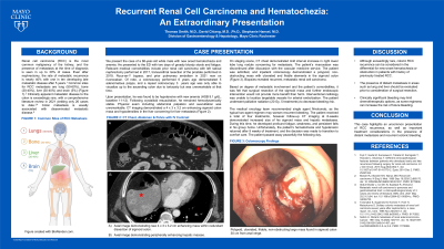
Figure: A) CT Abdomen & Pelvis with contrast demonstrating 4 x 3 x 3.2 cm enhancing mass in the sigmoid colon. B) Endoscopic imaging of non-obstructive, ulcerated, and friable mass in the sigmoid colon. C) Sigmoid mass biopsy demonstrating recurrent metastatic clear cell renal carcinoma, H&E stain. D) Sigmoid mass biopsy, KRT-CAM-5.2 stain.
Disclosures:
Stephanie Hansel indicated no relevant financial relationships.
David Chiang indicated no relevant financial relationships.
Thomas Smith indicated no relevant financial relationships.
Stephanie Hansel, MD, MS1, David Chiang, MD, PhD1, Thomas Smith, MD2. P0619 - Recurrent Renal Cell Carcinoma and Hematochezia: An Extraordinary Presentation, ACG 2023 Annual Scientific Meeting Abstracts. Vancouver, BC, Canada: American College of Gastroenterology.
