Sunday Poster Session
Category: GI Bleeding
P0620 - Hematochezia in a Patient With Hepatocellular Carcinoma: A Rectal Dieulafoy's Lesion Bleed
Sunday, October 22, 2023
3:30 PM - 7:00 PM PT
Location: Exhibit Hall

Has Audio

Riley Shumard
University of Kentucky
Lexington, KY
Presenting Author(s)
Angelica L. Solomon, MS, Riley Shumard, , Naba Saeed, MBBS
University of Kentucky, Lexington, KY
Introduction: A Dieulafoy’s lesion (DL) is a submucosal, dilated vascular lesion that can protrude through a small mucosal defect in the gastrointestinal tract, resulting in significant bleeding. Endoscopy has a diagnostic yield of 70%. Diagnostic criteria are: (I) active arterial bleeding from small (< 3mm) mucosal defects, with no ulceration or erosion of the mucosa; (II) visual evidence of raised artery; (III) blood clots with visible tiny point of mucosal attachments. DLs are not easily visible unless they are actively bleeding, leading to difficulty in establishing the diagnosis. Sometimes they may require CTA and IR embolization if they are unable to be endoscopically localized. They have been reported to account for up to 10% of GI bleeds. The most common site is the lesser curvature of the stomach (80-95%), but they can be found anywhere in the GI tract.
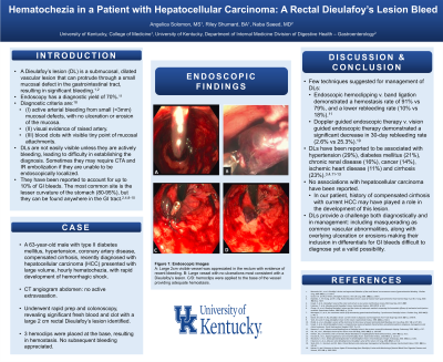
Case Description/Methods: A 63-year-old male with type II diabetes mellitus, hypertension, coronary artery disease, compensated cirrhosis, recently diagnosed with hepatocellular carcinoma (HCC) presented with large volume, hourly hematochezia, with rapid development of hemorrhagic shock. CT angiogram abdomen: no active extravasation. He underwent rapid prep and colonoscopy, revealing significant fresh blood and clot. Subsequently a large 2 cm rectal Dieulafoy's lesion was found. 3 hemoclips were placed at the base, resulting in hemostasis, and he did not have any subsequent bleeding.
Discussion: There are a few techniques suggested for management of Dieulafoy’s lesions. A comparison of endoscopic hemoclipping to band ligation demonstrated a hemostasis rate of 91% vs 79%, and a lower rebleeding rate (10% vs 18%). Another recent study compared doppler guided endoscopic therapy compared to vision guided endoscopic therapy and demonstrated a significant decrease in 30-day rebleeding rate (2.6% vs 25.3%). Our patient did well with hemoclips, without evidence of rebleeding.
DLs have been reported to be associated with Hypertension (29%), diabetes mellitus (21%), chronic renal disease (16%), cancer (14%), ischemic heart disease (11%) and cirrhosis (23%). There have been reports of gastric cancer appearing to be a dieulefoy lesion on initial presentation. No associations with hepatocellular carcinoma have been reported. In our patient, his history of compensated cirrhosis with current HCC may have played a role in the development of this lesion.

Disclosures:
Angelica L. Solomon, MS, Riley Shumard, , Naba Saeed, MBBS. P0620 - Hematochezia in a Patient With Hepatocellular Carcinoma: A Rectal Dieulafoy's Lesion Bleed, ACG 2023 Annual Scientific Meeting Abstracts. Vancouver, BC, Canada: American College of Gastroenterology.
University of Kentucky, Lexington, KY
Introduction: A Dieulafoy’s lesion (DL) is a submucosal, dilated vascular lesion that can protrude through a small mucosal defect in the gastrointestinal tract, resulting in significant bleeding. Endoscopy has a diagnostic yield of 70%. Diagnostic criteria are: (I) active arterial bleeding from small (< 3mm) mucosal defects, with no ulceration or erosion of the mucosa; (II) visual evidence of raised artery; (III) blood clots with visible tiny point of mucosal attachments. DLs are not easily visible unless they are actively bleeding, leading to difficulty in establishing the diagnosis. Sometimes they may require CTA and IR embolization if they are unable to be endoscopically localized. They have been reported to account for up to 10% of GI bleeds. The most common site is the lesser curvature of the stomach (80-95%), but they can be found anywhere in the GI tract.
Case Description/Methods: A 63-year-old male with type II diabetes mellitus, hypertension, coronary artery disease, compensated cirrhosis, recently diagnosed with hepatocellular carcinoma (HCC) presented with large volume, hourly hematochezia, with rapid development of hemorrhagic shock. CT angiogram abdomen: no active extravasation. He underwent rapid prep and colonoscopy, revealing significant fresh blood and clot. Subsequently a large 2 cm rectal Dieulafoy's lesion was found. 3 hemoclips were placed at the base, resulting in hemostasis, and he did not have any subsequent bleeding.
Discussion: There are a few techniques suggested for management of Dieulafoy’s lesions. A comparison of endoscopic hemoclipping to band ligation demonstrated a hemostasis rate of 91% vs 79%, and a lower rebleeding rate (10% vs 18%). Another recent study compared doppler guided endoscopic therapy compared to vision guided endoscopic therapy and demonstrated a significant decrease in 30-day rebleeding rate (2.6% vs 25.3%). Our patient did well with hemoclips, without evidence of rebleeding.
DLs have been reported to be associated with Hypertension (29%), diabetes mellitus (21%), chronic renal disease (16%), cancer (14%), ischemic heart disease (11%) and cirrhosis (23%). There have been reports of gastric cancer appearing to be a dieulefoy lesion on initial presentation. No associations with hepatocellular carcinoma have been reported. In our patient, his history of compensated cirrhosis with current HCC may have played a role in the development of this lesion.

Figure: A - a large 2cm visible vessel was appreciated in the rectum with evidence of recent bleeding B - large vessel with no ulceration most consistent with a Dieulafoy's lesion C - hemoclips were applied to the base of the vessel providing adequate hemostasis
Disclosures:
Angelica Solomon indicated no relevant financial relationships.
Riley Shumard indicated no relevant financial relationships.
Naba Saeed indicated no relevant financial relationships.
Angelica L. Solomon, MS, Riley Shumard, , Naba Saeed, MBBS. P0620 - Hematochezia in a Patient With Hepatocellular Carcinoma: A Rectal Dieulafoy's Lesion Bleed, ACG 2023 Annual Scientific Meeting Abstracts. Vancouver, BC, Canada: American College of Gastroenterology.
