Sunday Poster Session
Category: GI Bleeding
P0625 - Renal Cell Carcinoma of the Stomach Disguised as Hematemesis
Sunday, October 22, 2023
3:30 PM - 7:00 PM PT
Location: Exhibit Hall

Has Audio
- LW
Lauren Walters, BA
Creighton University School of Medicine
Omaha, NE
Presenting Author(s)
Lauren Walters, BA1, Suhail Sidhu, BS1, T. Rees Checketts, DO1, Mohammed Qasswal, MD2, Sahil Mullick, MD2, Sandeep Mukherjee, MD2
1Creighton University School of Medicine, Omaha, NE; 2Creighton University, Omaha, NE
Introduction: Renal cell carcinoma (RCC) is the leading cause of kidney cancer and most commonly metastasizes to the lungs, bones, and lymph nodes. Metastases to the stomach are rare, occurring in only 0.2% of cases. Here, we describe a case of gastric metastases from RCC presenting as an upper gastrointestinal (GI) bleed three years after the patient was thought to have achieved remission.
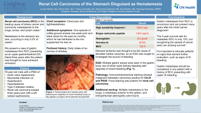
Case Description/Methods: A 79 year-old-male with a past medical history of a remote aortic valve replacement, two myocardial infarctions, hypertension, type II diabetes mellitus, and renal cell carcinoma treated three years prior with a left radical nephrectomy presented for chest pain and lightheadedness. His initial workup revealed no acute cardiopulmonary process on electrocardiogram, but an elevated high sensitivity troponin and brain natriuretic peptide of 538.2ng/L and 1,621pg/ml, respectively. He was also found to have new onset anemia with a hemoglobin of 8.2gm/dl, hematocrit of 26.8%, and an increased mean corpuscular volume of 102fl. Upon further questioning, the patient admitted to having one episode of black, tarry vomit one week prior and black stools for the past six months. He attributed his abnormal bowel movements to the iron supplement he had been taking since his nephrectomy. He also admitted to drinking six ounces of whiskey daily. Rectal exam showed small specks of black stool with no bright red blood. Cardiology determined the patient’s cardiac enzymes were elevated due to demand ischemia, so an esophagogastroduodenoscopy (EGD) was sought to investigate the source of a suspected upper GI bleed. Multiple gastric polyps were visualized in the gastric body, two of which were actively bleeding with stigmata of recent bleeding (Fig. 1). The polyps were clipped and biopsies were sent to pathology. Immunohistochemical staining showed malignant metastatic carcinoma positive for CA-IX and PAX8. Focal staining was positive for RCC. Additional workup demonstrated multiple metastases to the lungs, a metastasis anterior to the spleen, and retroperitoneal adenopathy.
Discussion: The development of gastric metastases from RCC is uncommon and can present several years after the initial cancer diagnosis. The 5-year survival rate for metastatic RCC is only 12%, but recognizing the spread of cancer early can prolong survival. Therefore, it is important to educate patients on signs that warrant further evaluation and to consider gastric metastases in any patient presenting with upper GI bleeding and a history of RCC.

Disclosures:
Lauren Walters, BA1, Suhail Sidhu, BS1, T. Rees Checketts, DO1, Mohammed Qasswal, MD2, Sahil Mullick, MD2, Sandeep Mukherjee, MD2. P0625 - Renal Cell Carcinoma of the Stomach Disguised as Hematemesis, ACG 2023 Annual Scientific Meeting Abstracts. Vancouver, BC, Canada: American College of Gastroenterology.
1Creighton University School of Medicine, Omaha, NE; 2Creighton University, Omaha, NE
Introduction: Renal cell carcinoma (RCC) is the leading cause of kidney cancer and most commonly metastasizes to the lungs, bones, and lymph nodes. Metastases to the stomach are rare, occurring in only 0.2% of cases. Here, we describe a case of gastric metastases from RCC presenting as an upper gastrointestinal (GI) bleed three years after the patient was thought to have achieved remission.
Case Description/Methods: A 79 year-old-male with a past medical history of a remote aortic valve replacement, two myocardial infarctions, hypertension, type II diabetes mellitus, and renal cell carcinoma treated three years prior with a left radical nephrectomy presented for chest pain and lightheadedness. His initial workup revealed no acute cardiopulmonary process on electrocardiogram, but an elevated high sensitivity troponin and brain natriuretic peptide of 538.2ng/L and 1,621pg/ml, respectively. He was also found to have new onset anemia with a hemoglobin of 8.2gm/dl, hematocrit of 26.8%, and an increased mean corpuscular volume of 102fl. Upon further questioning, the patient admitted to having one episode of black, tarry vomit one week prior and black stools for the past six months. He attributed his abnormal bowel movements to the iron supplement he had been taking since his nephrectomy. He also admitted to drinking six ounces of whiskey daily. Rectal exam showed small specks of black stool with no bright red blood. Cardiology determined the patient’s cardiac enzymes were elevated due to demand ischemia, so an esophagogastroduodenoscopy (EGD) was sought to investigate the source of a suspected upper GI bleed. Multiple gastric polyps were visualized in the gastric body, two of which were actively bleeding with stigmata of recent bleeding (Fig. 1). The polyps were clipped and biopsies were sent to pathology. Immunohistochemical staining showed malignant metastatic carcinoma positive for CA-IX and PAX8. Focal staining was positive for RCC. Additional workup demonstrated multiple metastases to the lungs, a metastasis anterior to the spleen, and retroperitoneal adenopathy.
Discussion: The development of gastric metastases from RCC is uncommon and can present several years after the initial cancer diagnosis. The 5-year survival rate for metastatic RCC is only 12%, but recognizing the spread of cancer early can prolong survival. Therefore, it is important to educate patients on signs that warrant further evaluation and to consider gastric metastases in any patient presenting with upper GI bleeding and a history of RCC.

Figure: Fig. 1. Pedunculated and sessile polyp with bleeding and stigmata of recent bleeding found on the greater curvature of the stomach.
Disclosures:
Lauren Walters indicated no relevant financial relationships.
Suhail Sidhu indicated no relevant financial relationships.
T. Rees Checketts indicated no relevant financial relationships.
Mohammed Qasswal indicated no relevant financial relationships.
Sahil Mullick indicated no relevant financial relationships.
Sandeep Mukherjee: Dynamed Plus – Section editor for Hepatology. Gilead – Speakers Bureau.
Lauren Walters, BA1, Suhail Sidhu, BS1, T. Rees Checketts, DO1, Mohammed Qasswal, MD2, Sahil Mullick, MD2, Sandeep Mukherjee, MD2. P0625 - Renal Cell Carcinoma of the Stomach Disguised as Hematemesis, ACG 2023 Annual Scientific Meeting Abstracts. Vancouver, BC, Canada: American College of Gastroenterology.
