Sunday Poster Session
Category: GI Bleeding
P0633 - Pernicious Pouch of Problems: A Challenging Case of Massive Hemorrhage Secondary to Jejunal Diverticular Bleeding
Sunday, October 22, 2023
3:30 PM - 7:00 PM PT
Location: Exhibit Hall

Has Audio
.jpg)
Tyler J. Reed, MD, BA
Naval Medical Center
Portsmouth, VA
Presenting Author(s)
Tyler Reed, MD, BA, Allison Bush, MD, Nicholas Nelson, MD
Naval Medical Center, Portsmouth, VA
Introduction: Small bowel (SB) diverticulosis is an uncommon condition with jejunal diverticulosis being particularly rare, estimated to occur in less than 1% of people. SB diverticular bleeding can be difficult to identify and treat. We present a case of a severe jejunal diverticular hemorrhage requiring massive transfusion protocol with diagnostic and therapeutic challenges.
Case Description/Methods: A 57-year-old female with a history of polymyositis and NSAID use presented to the emergency department with one day of hematemesis, rectal bleeding and syncope. Complete blood cell count revealed a hemoglobin of 5.5 g/dL. She was admitted to the intensive care unit. Computed tomography angiography (CTA) of the abdomen and pelvis was unrevealing. Esophagogastroduodenoscopy (EGD) and demonstrated SB diverticula but no source of blood loss. Interventional Radiology (IR) performed directed angiography which showed no evidence of arterial extravasation. Repeat EGD and push enteroscopy again showed multiple SB diverticula without active bleeding, clot or visible vessels. Colonoscopy demonstrated no diverticula or active bleeding. She was transfused 19 units of packed red blood cells over the hospitalization with ongoing hematochezia. After another episode of large-volume hematochezia, repeat urgent CTA demonstrated several diverticula including a 1.8 cm jejunal diverticula with active arterial bleeding. Due the location and risk of ischemia, IR did not recommend embolization. A repeat EGD and push enteroscopy revealed a large jejunal diverticulum with a non-bleeding visible vessel treated with epinephrine and placement of three clips. The bleeding resolved and she was discharged home.
Discussion: Risk factors for SB diverticulosis include intestinal dysmotility syndromes and visceral myopathy. The duodenum is the most common location for SB diverticulosis with jejunal diverticulosis being the least common location. SB diverticulosis is usually asymptomatic, but patients may report early satiety, abdominal discomfort or bloating. Severe bleeding from jejunal diverticulosis is rare. The jejunum is a location that can be challenging to diagnose and treat both endoscopically and with IR. This case demonstrates a rare yet potentially life-threatening complication of jejunal diverticulosis with massive hemorrhage that evaded multiple diagnostic evaluations, that was ultimately treated endoscopically without long term complications.

Disclosures:
Tyler Reed, MD, BA, Allison Bush, MD, Nicholas Nelson, MD. P0633 - Pernicious Pouch of Problems: A Challenging Case of Massive Hemorrhage Secondary to Jejunal Diverticular Bleeding, ACG 2023 Annual Scientific Meeting Abstracts. Vancouver, BC, Canada: American College of Gastroenterology.
Naval Medical Center, Portsmouth, VA
Introduction: Small bowel (SB) diverticulosis is an uncommon condition with jejunal diverticulosis being particularly rare, estimated to occur in less than 1% of people. SB diverticular bleeding can be difficult to identify and treat. We present a case of a severe jejunal diverticular hemorrhage requiring massive transfusion protocol with diagnostic and therapeutic challenges.
Case Description/Methods: A 57-year-old female with a history of polymyositis and NSAID use presented to the emergency department with one day of hematemesis, rectal bleeding and syncope. Complete blood cell count revealed a hemoglobin of 5.5 g/dL. She was admitted to the intensive care unit. Computed tomography angiography (CTA) of the abdomen and pelvis was unrevealing. Esophagogastroduodenoscopy (EGD) and demonstrated SB diverticula but no source of blood loss. Interventional Radiology (IR) performed directed angiography which showed no evidence of arterial extravasation. Repeat EGD and push enteroscopy again showed multiple SB diverticula without active bleeding, clot or visible vessels. Colonoscopy demonstrated no diverticula or active bleeding. She was transfused 19 units of packed red blood cells over the hospitalization with ongoing hematochezia. After another episode of large-volume hematochezia, repeat urgent CTA demonstrated several diverticula including a 1.8 cm jejunal diverticula with active arterial bleeding. Due the location and risk of ischemia, IR did not recommend embolization. A repeat EGD and push enteroscopy revealed a large jejunal diverticulum with a non-bleeding visible vessel treated with epinephrine and placement of three clips. The bleeding resolved and she was discharged home.
Discussion: Risk factors for SB diverticulosis include intestinal dysmotility syndromes and visceral myopathy. The duodenum is the most common location for SB diverticulosis with jejunal diverticulosis being the least common location. SB diverticulosis is usually asymptomatic, but patients may report early satiety, abdominal discomfort or bloating. Severe bleeding from jejunal diverticulosis is rare. The jejunum is a location that can be challenging to diagnose and treat both endoscopically and with IR. This case demonstrates a rare yet potentially life-threatening complication of jejunal diverticulosis with massive hemorrhage that evaded multiple diagnostic evaluations, that was ultimately treated endoscopically without long term complications.

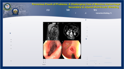
Figure: A) CTA abdomen and pelvis with contrast, coronal view, demonstrating approximately 1.8cm jejunal diverticulum
B) CTA abdomen and pelvis with contrast, axial view, arterial phase, concerning for active extravasation
C,D) EGD and push enteroscopy with visible vessel inside large jejunal diverticulum
B) CTA abdomen and pelvis with contrast, axial view, arterial phase, concerning for active extravasation
C,D) EGD and push enteroscopy with visible vessel inside large jejunal diverticulum
Disclosures:
Tyler Reed indicated no relevant financial relationships.
Allison Bush indicated no relevant financial relationships.
Nicholas Nelson indicated no relevant financial relationships.
Tyler Reed, MD, BA, Allison Bush, MD, Nicholas Nelson, MD. P0633 - Pernicious Pouch of Problems: A Challenging Case of Massive Hemorrhage Secondary to Jejunal Diverticular Bleeding, ACG 2023 Annual Scientific Meeting Abstracts. Vancouver, BC, Canada: American College of Gastroenterology.
