Sunday Poster Session
Category: GI Bleeding
P0636 - In the Shadows of the Veil: Unraveling the Enigma of Rectal Bleeding and Proctalgy
Sunday, October 22, 2023
3:30 PM - 7:00 PM PT
Location: Exhibit Hall


Lizeth Cifuentes, MD
University of Pittsburgh Medical Center
Pittsburgh, PA
Presenting Author(s)
Lizeth Cifuentes, MD1, Ivan Del pozo, MD2
1University of Pittsburgh Medical Center, Pittsburgh, PA; 2Hospital Universitario Rey Juan Carlos, Madrid, Madrid, Spain
Introduction: A 39-year-old man visited the clinic with complaints of rectal bleeding and proctalgia persisting for the past three months. He reported no significant medical or surgical history, and denied the use of alcohol or tobacco. He confirmed being sexually active with his wife and denied engaging in anal-receptive sexual activities. A rectal examination revealed the presence of a 1.0 cm round-shaped, polypoid mass located approximately 3cm from the anal verge. No signs of inguinal lymphadenopathy were noted.
Case Description/Methods: During the rectal examination, a 1.0 cm round-shaped, polypoid mass was discovered approximately 3cm from the anal verge. No inguinal lymphadenopathy was observed.
Discussion: The histopathological analysis of the specimen confirms the presence of lymphangioma. Lymphangiomas typically manifest as submucosal polypoid growths that generally do not cause symptoms. However, in rare cases, patients may experience abdominal pain, discomfort, or rectal bleeding. Lymphangioma is characterized by clusters of translucent small vesicles, which can range in color from clear to dark. These vesicles are formed by cystically dilated lymphatic channels embedded in a myxoid stroma that may contain lymphocytes. The vascular channels within lymphangiomas typically contain eosinophilic material but lack red blood cells.
Lymphangioma circumscripum of the rectum is an infrequent hamartomatous malformation that not only affects the skin but can also spread to the mucosal tissue in some instances. It is an extremely rare condition, with fewer than 100 cases reported in the literature. Lymphangioma circumscriptum represents a developmental abnormality of lymphatics in the deep dermal and subcutaneous layers. It can occur as either a congenital anomaly or an acquired form arising from previously normal lymphatics. No malignant variant (lymphangiosarcoma), either histologically or clinically, has been reported.
The diagnosis of lymphangioma is typically confirmed through histopathological examination of the lesion, which may resemble other conditions such as polyps (adenomas), fistulas, cancer, hemorrhoids, inflammatory diseases, or infectious collections.
The standard treatment for lymphangioma is surgical excision.

Disclosures:
Lizeth Cifuentes, MD1, Ivan Del pozo, MD2. P0636 - In the Shadows of the Veil: Unraveling the Enigma of Rectal Bleeding and Proctalgy, ACG 2023 Annual Scientific Meeting Abstracts. Vancouver, BC, Canada: American College of Gastroenterology.
1University of Pittsburgh Medical Center, Pittsburgh, PA; 2Hospital Universitario Rey Juan Carlos, Madrid, Madrid, Spain
Introduction: A 39-year-old man visited the clinic with complaints of rectal bleeding and proctalgia persisting for the past three months. He reported no significant medical or surgical history, and denied the use of alcohol or tobacco. He confirmed being sexually active with his wife and denied engaging in anal-receptive sexual activities. A rectal examination revealed the presence of a 1.0 cm round-shaped, polypoid mass located approximately 3cm from the anal verge. No signs of inguinal lymphadenopathy were noted.
Case Description/Methods: During the rectal examination, a 1.0 cm round-shaped, polypoid mass was discovered approximately 3cm from the anal verge. No inguinal lymphadenopathy was observed.
Discussion: The histopathological analysis of the specimen confirms the presence of lymphangioma. Lymphangiomas typically manifest as submucosal polypoid growths that generally do not cause symptoms. However, in rare cases, patients may experience abdominal pain, discomfort, or rectal bleeding. Lymphangioma is characterized by clusters of translucent small vesicles, which can range in color from clear to dark. These vesicles are formed by cystically dilated lymphatic channels embedded in a myxoid stroma that may contain lymphocytes. The vascular channels within lymphangiomas typically contain eosinophilic material but lack red blood cells.
Lymphangioma circumscripum of the rectum is an infrequent hamartomatous malformation that not only affects the skin but can also spread to the mucosal tissue in some instances. It is an extremely rare condition, with fewer than 100 cases reported in the literature. Lymphangioma circumscriptum represents a developmental abnormality of lymphatics in the deep dermal and subcutaneous layers. It can occur as either a congenital anomaly or an acquired form arising from previously normal lymphatics. No malignant variant (lymphangiosarcoma), either histologically or clinically, has been reported.
The diagnosis of lymphangioma is typically confirmed through histopathological examination of the lesion, which may resemble other conditions such as polyps (adenomas), fistulas, cancer, hemorrhoids, inflammatory diseases, or infectious collections.
The standard treatment for lymphangioma is surgical excision.

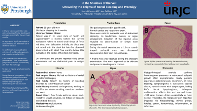
Figure: Figure 1. A) Panoramic view. Cystically dilated lymphatic channels. The stroma contains lymphocytes. B) The spaces are lined by flat endothelium, containing eosinophilic fluid without red blood cells. HE
Disclosures:
Lizeth Cifuentes indicated no relevant financial relationships.
Ivan Del pozo indicated no relevant financial relationships.
Lizeth Cifuentes, MD1, Ivan Del pozo, MD2. P0636 - In the Shadows of the Veil: Unraveling the Enigma of Rectal Bleeding and Proctalgy, ACG 2023 Annual Scientific Meeting Abstracts. Vancouver, BC, Canada: American College of Gastroenterology.
