Sunday Poster Session
Category: GI Bleeding
P0638 - Gastrointestinal Tract Complications From Iododerma
Sunday, October 22, 2023
3:30 PM - 7:00 PM PT
Location: Exhibit Hall

Has Audio

Patricia Miguez Arosemena, MD
Mount Sinai West and Morningside
New York, NY
Presenting Author(s)
Patricia Miguez Arosemena, MD1, Andre Khazak, DO2, Angad Uberoi, MD1, Shanique Wilson Noack, MD1, Kimberly Cavaliere, MD3
1Mount Sinai West and Morningside, New York, NY; 2Mount Sinai Beth Israel, New York, NY; 3Mount Sinai Morningside-West, New York, NY
Introduction: Iododerma is a rare and potentially serious dermatosis that develops as a delayed hypersensitive reaction to iodinated compounds, often occurring in patients with renal insufficiency. Cutaneous presentation can vary but includes nodular eruption, acneiform, pustular, and hemorrhagic bullae. Symptoms typically start 2-3 days following exposure and resolve within 2-6 weeks. Extracutaneous manifestations include conjunctivitis, salivary gland swelling, vasculitis, and respiratory compromise. We present a case of a patient with iododerma and gastrointestinal manifestations.
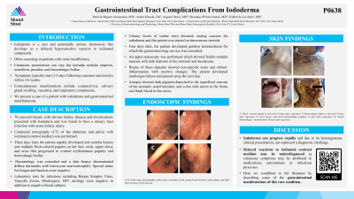
Case Description/Methods: A 78-year-old female with chronic kidney disease and diverticulosis presented with hematuria and was found to have a urinary tract infection with acute kidney injury. Computed tomography (CT) of the abdomen and pelvis with iodinated contrast medium was performed. Three days later, the patient rapidly developed oral nodular lesions and multiple flesh-colored papules on her face, neck, upper chest, and arms that progressed to crusted erythematous papules and hemorrhagic bullae. Dermatology was consulted and a skin biopsy demonstrated diffuse dermatitis with histiocytes and neutrophils. Special stains for fungus and bacteria were negative. Laboratory tests for infections including Herpes Simplex Virus, Varicella Zoster, Monkeypox, HIV serology were negative in addition to negative blood cultures. Urinary levels of iodine were elevated, raising concern for iododerma and the patient was started on intravenous steroids. Four days later, the patient developed painless hematochezia for which the gastroenterology service was consulted. An upper endoscopy was performed which showed friable nodular mucosa with dark deposits in the stomach and duodenum. Biopsy of these deposits showed non-specific acute and chronic inflammation with reactive changes. The patient developed multiorgan failure and passed away the next day. Autopsy showed dark pigment deposited in the superficial mucosa of the stomach, small intestine, and colon with ulcers in the ileum and frank blood in the colon.
Discussion: Iododerma can progress rapidly and due to its heterogeneous clinical presentation, can represent a diagnostic challenge. Delayed reactions to iodinated contrast medium may be underdiagnosed as cutaneous symptoms may be attributed to medications, autoimmune or infectious processes. Here we contribute to the literature by describing some of the gastrointestinal manifestations of this rare condition.

Disclosures:
Patricia Miguez Arosemena, MD1, Andre Khazak, DO2, Angad Uberoi, MD1, Shanique Wilson Noack, MD1, Kimberly Cavaliere, MD3. P0638 - Gastrointestinal Tract Complications From Iododerma, ACG 2023 Annual Scientific Meeting Abstracts. Vancouver, BC, Canada: American College of Gastroenterology.
1Mount Sinai West and Morningside, New York, NY; 2Mount Sinai Beth Israel, New York, NY; 3Mount Sinai Morningside-West, New York, NY
Introduction: Iododerma is a rare and potentially serious dermatosis that develops as a delayed hypersensitive reaction to iodinated compounds, often occurring in patients with renal insufficiency. Cutaneous presentation can vary but includes nodular eruption, acneiform, pustular, and hemorrhagic bullae. Symptoms typically start 2-3 days following exposure and resolve within 2-6 weeks. Extracutaneous manifestations include conjunctivitis, salivary gland swelling, vasculitis, and respiratory compromise. We present a case of a patient with iododerma and gastrointestinal manifestations.
Case Description/Methods: A 78-year-old female with chronic kidney disease and diverticulosis presented with hematuria and was found to have a urinary tract infection with acute kidney injury. Computed tomography (CT) of the abdomen and pelvis with iodinated contrast medium was performed. Three days later, the patient rapidly developed oral nodular lesions and multiple flesh-colored papules on her face, neck, upper chest, and arms that progressed to crusted erythematous papules and hemorrhagic bullae. Dermatology was consulted and a skin biopsy demonstrated diffuse dermatitis with histiocytes and neutrophils. Special stains for fungus and bacteria were negative. Laboratory tests for infections including Herpes Simplex Virus, Varicella Zoster, Monkeypox, HIV serology were negative in addition to negative blood cultures. Urinary levels of iodine were elevated, raising concern for iododerma and the patient was started on intravenous steroids. Four days later, the patient developed painless hematochezia for which the gastroenterology service was consulted. An upper endoscopy was performed which showed friable nodular mucosa with dark deposits in the stomach and duodenum. Biopsy of these deposits showed non-specific acute and chronic inflammation with reactive changes. The patient developed multiorgan failure and passed away the next day. Autopsy showed dark pigment deposited in the superficial mucosa of the stomach, small intestine, and colon with ulcers in the ileum and frank blood in the colon.
Discussion: Iododerma can progress rapidly and due to its heterogeneous clinical presentation, can represent a diagnostic challenge. Delayed reactions to iodinated contrast medium may be underdiagnosed as cutaneous symptoms may be attributed to medications, autoimmune or infectious processes. Here we contribute to the literature by describing some of the gastrointestinal manifestations of this rare condition.

Figure: A-D. Endoscopic photography of the lesser curvature of the stomach and incisura with nodules and dark discoloration of the mucosa.
E. Flesh-colored papules in the neck (Three days after exposure).
F. Hemorrhagic bullae in the neck (Five days after exposure).
G. Oral ulcers and facial hemorrhagic papules (Five days after exposure).
H. Facial hemorrhagic crusted lesions (Eight days after exposure).
E. Flesh-colored papules in the neck (Three days after exposure).
F. Hemorrhagic bullae in the neck (Five days after exposure).
G. Oral ulcers and facial hemorrhagic papules (Five days after exposure).
H. Facial hemorrhagic crusted lesions (Eight days after exposure).
Disclosures:
Patricia Miguez Arosemena indicated no relevant financial relationships.
Andre Khazak indicated no relevant financial relationships.
Angad Uberoi indicated no relevant financial relationships.
Shanique Wilson Noack indicated no relevant financial relationships.
Kimberly Cavaliere indicated no relevant financial relationships.
Patricia Miguez Arosemena, MD1, Andre Khazak, DO2, Angad Uberoi, MD1, Shanique Wilson Noack, MD1, Kimberly Cavaliere, MD3. P0638 - Gastrointestinal Tract Complications From Iododerma, ACG 2023 Annual Scientific Meeting Abstracts. Vancouver, BC, Canada: American College of Gastroenterology.
