Sunday Poster Session
Category: GI Bleeding
P0639 - A Slow Bleed: A Case Report of a Large Colonic Arteriovenous Malformations in a 23-Year-Old Male
Sunday, October 22, 2023
3:30 PM - 7:00 PM PT
Location: Exhibit Hall

Has Audio
- MB
Matthew D. Brockway, DO
MountainView Hospital
Las Vegas, NV
Presenting Author(s)
Matthew D. Brockway, DO, Victoria Diaz, MD, Reed Hansen, DO, Milliejoan Mongalo, MD, Jordan Valenta, DO, Homayon Iraninezhad, DO
MountainView Hospital, Las Vegas, NV
Introduction: Arteriovenous malformations (AVM) are irregular connections between arteries and veins, causing hypoxia and hemorrhage. AVMs are usually solitarily lesions or may occur as a syndrome. AVMs are rare lesions that comprise 0.8 – 3% of lower gastrointestinal bleeds and are very rare in patients under 50 years. This case is a unique presentation and the only one that we know of a large AVM involving the ascending, and half of the transverse colon, and being circumferential throughout.
Case Description/Methods: A 23-year-old male presented to the emergency department with weakness, fatigue, and bright red blood per rectum intermittently ongoing since high school. The patient’s last episode was a month ago. The patient reports a colonoscopy in 2020 showing no significant findings. The patient uses tobacco, alcohol, and occasionally cocaine. Initial labs were significant for Hgb 3.9, WBC count 2.5, MCV 56, PT 11.9, BMP within normal limits, iron 8, iron saturation 2, ferritin < 1, reticulocyte count of 1.3, and B12, folate and liver function tests within normal limits.
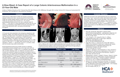
Esophagogastroduodenoscopy (EGD) showed no bleeding. Colonoscopy showed enlarged veins in the right colon with one oozing blood (Image A, B, C). A resolution clip was placed with no further bleeding noted. CT abdomen and pelvis with IV contrast was ordered to assess the enlarged vasculature noted on colonoscopy. This showed the ascending colon supplied by multiple feeding branches off the superior mesenteric artery and early contrast opacification of the superior mesenteric vein consistent with AVM (Image D, E). The patient had no findings consistent with Hereditary Hemorrhagic Telangiectasia (HHT). The patient was taken to vascular surgery and had the additional proximal vessels attached to the SMA ligated. The patient had no signs of bleeding or ischemic bowel and was told to follow up in a month. A month later, the patient underwent embolization of 3 more vessels (Image F). The patient has had minimal bleeding with a stable hemoglobin since surgery.
Discussion: AVMs that are found in the gastrointestinal tract are categorized into 3 types. Type 1 lesions are solitary right-sided lesions usually found in the elderly. Type 2 lesions are large, in the small bowel, congenital, and found in younger patients. Type 3 lesions are punctate angiomas associated with HHT. Treatment of these lesions can be endoscopic, surgical, or embolization. It’s important to keep these lesions on the differential and not default them to elderly patients.

Disclosures:
Matthew D. Brockway, DO, Victoria Diaz, MD, Reed Hansen, DO, Milliejoan Mongalo, MD, Jordan Valenta, DO, Homayon Iraninezhad, DO. P0639 - A Slow Bleed: A Case Report of a Large Colonic Arteriovenous Malformations in a 23-Year-Old Male, ACG 2023 Annual Scientific Meeting Abstracts. Vancouver, BC, Canada: American College of Gastroenterology.
MountainView Hospital, Las Vegas, NV
Introduction: Arteriovenous malformations (AVM) are irregular connections between arteries and veins, causing hypoxia and hemorrhage. AVMs are usually solitarily lesions or may occur as a syndrome. AVMs are rare lesions that comprise 0.8 – 3% of lower gastrointestinal bleeds and are very rare in patients under 50 years. This case is a unique presentation and the only one that we know of a large AVM involving the ascending, and half of the transverse colon, and being circumferential throughout.
Case Description/Methods: A 23-year-old male presented to the emergency department with weakness, fatigue, and bright red blood per rectum intermittently ongoing since high school. The patient’s last episode was a month ago. The patient reports a colonoscopy in 2020 showing no significant findings. The patient uses tobacco, alcohol, and occasionally cocaine. Initial labs were significant for Hgb 3.9, WBC count 2.5, MCV 56, PT 11.9, BMP within normal limits, iron 8, iron saturation 2, ferritin < 1, reticulocyte count of 1.3, and B12, folate and liver function tests within normal limits.
Esophagogastroduodenoscopy (EGD) showed no bleeding. Colonoscopy showed enlarged veins in the right colon with one oozing blood (Image A, B, C). A resolution clip was placed with no further bleeding noted. CT abdomen and pelvis with IV contrast was ordered to assess the enlarged vasculature noted on colonoscopy. This showed the ascending colon supplied by multiple feeding branches off the superior mesenteric artery and early contrast opacification of the superior mesenteric vein consistent with AVM (Image D, E). The patient had no findings consistent with Hereditary Hemorrhagic Telangiectasia (HHT). The patient was taken to vascular surgery and had the additional proximal vessels attached to the SMA ligated. The patient had no signs of bleeding or ischemic bowel and was told to follow up in a month. A month later, the patient underwent embolization of 3 more vessels (Image F). The patient has had minimal bleeding with a stable hemoglobin since surgery.
Discussion: AVMs that are found in the gastrointestinal tract are categorized into 3 types. Type 1 lesions are solitary right-sided lesions usually found in the elderly. Type 2 lesions are large, in the small bowel, congenital, and found in younger patients. Type 3 lesions are punctate angiomas associated with HHT. Treatment of these lesions can be endoscopic, surgical, or embolization. It’s important to keep these lesions on the differential and not default them to elderly patients.

Figure: Figure: Image A, B, and C. Endoscopic imaging showing large circumferential AVM with an area of bleeding and status post resolution clip. Image D, E. Coronal computed tomography angiography (CTA) image showing large AVM coming from the superior mesenteric artery going to the ascending colon. Image F. Coronal CTA image showing the superior mesenteric artery AVM status post ligation and embolization.
Disclosures:
Matthew Brockway indicated no relevant financial relationships.
Victoria Diaz indicated no relevant financial relationships.
Reed Hansen indicated no relevant financial relationships.
Milliejoan Mongalo indicated no relevant financial relationships.
Jordan Valenta indicated no relevant financial relationships.
Homayon Iraninezhad indicated no relevant financial relationships.
Matthew D. Brockway, DO, Victoria Diaz, MD, Reed Hansen, DO, Milliejoan Mongalo, MD, Jordan Valenta, DO, Homayon Iraninezhad, DO. P0639 - A Slow Bleed: A Case Report of a Large Colonic Arteriovenous Malformations in a 23-Year-Old Male, ACG 2023 Annual Scientific Meeting Abstracts. Vancouver, BC, Canada: American College of Gastroenterology.
