Sunday Poster Session
Category: GI Bleeding
P0641 - When a Gastrointestinal Bleed Unveils a Surprising Diagnosis
Sunday, October 22, 2023
3:30 PM - 7:00 PM PT
Location: Exhibit Hall

Has Audio

Jose I. De Arrigunaga, DO
University of Texas Health Science Center at San Antonio
San Antonio, TX
Presenting Author(s)
Jose I.. De Arrigunaga, DO1, Umar Siddiqui, DO2, Theodore V.. Arevalo, MD1
1University of Texas Health Science Center at San Antonio, San Antonio, TX; 2University of Texas Health Science Center-San Antonio, San Antonio, TX
Introduction: Primary gastrointestinal lymphomas account for nearly 6-12% of all malignancies; the two most common subtypes are Diffuse Large B-Cell Lymphoma (DLBCL) and Mucosa Associated Lymphoid Tissue (MALToma). Most patients with gastrointestinal lymphomas are symptomatic and report abdominal pain, hematochezia, nausea, vomiting, fever, and weight loss. Bladder lymphomas, which account for less than 1% of all neoplasms, generally present with some constellation of urinary tract symptoms, fatigue, and weight loss. In this case, an old-aged male presented with a gastrointestinal bleed which led to the discovery of two primary gastrointestinal lymphomas with metastasis.
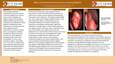
Case Description/Methods: An 85-year-old male with a past medical history of diverticulosis and internal hemorrhoids presented with melena for 6 months and one episode of hematochezia three days prior to admission. The patient denied weight loss, fevers, night sweats, fatigue, abdominal pain, rectal pain, hematuria, dysuria, urinary frequency, and suprapubic tenderness. The patient reported two previously unremarkable colonoscopies. Significant medications included aspirin. Family history showed no first-degree relatives with colon cancer. Abdominal exam was unremarkable, and a digital rectal exam was significant for an external hemorrhoid; however, there was no blood in the rectal vault. Hemoglobin was stable. Colonoscopy and esophagogastroduodenoscopy showed a rectal mass and erythematous, friable, mucosa in the stomach, respectively. Biopsy of the masses showed DLBCL in the rectum (Image A) and Helicobacter pylori-negative MALToma in the stomach (Image B). Bone marrow biopsy was negative. Magnetic Resonance Imaging of Abdomen and Pelvis was pursued and ultimately showed a bladder wall mass. A transurethral resection of the bladder tumor was performed and showed MALToma likely metastatic from the gastric MALToma.
Discussion: This case represents a rare scenario where two primary gastrointestinal lymphomas with metastasis to the bladder wall were unmasked in the setting of a gastrointestinal bleed and no additional symptoms. The absence of symptoms may lead certain healthcare providers to forego additional imaging. However, this case highlights the importance of imaging, and its role in the workup and staging of newly diagnosed gastrointestinal lymphoma to exclude involvement of other sites even in the absence of symptoms. To our knowledge, there are no prior cases in the literature that show bladder wall MALToma with little to no symptoms.

Disclosures:
Jose I.. De Arrigunaga, DO1, Umar Siddiqui, DO2, Theodore V.. Arevalo, MD1. P0641 - When a Gastrointestinal Bleed Unveils a Surprising Diagnosis, ACG 2023 Annual Scientific Meeting Abstracts. Vancouver, BC, Canada: American College of Gastroenterology.
1University of Texas Health Science Center at San Antonio, San Antonio, TX; 2University of Texas Health Science Center-San Antonio, San Antonio, TX
Introduction: Primary gastrointestinal lymphomas account for nearly 6-12% of all malignancies; the two most common subtypes are Diffuse Large B-Cell Lymphoma (DLBCL) and Mucosa Associated Lymphoid Tissue (MALToma). Most patients with gastrointestinal lymphomas are symptomatic and report abdominal pain, hematochezia, nausea, vomiting, fever, and weight loss. Bladder lymphomas, which account for less than 1% of all neoplasms, generally present with some constellation of urinary tract symptoms, fatigue, and weight loss. In this case, an old-aged male presented with a gastrointestinal bleed which led to the discovery of two primary gastrointestinal lymphomas with metastasis.
Case Description/Methods: An 85-year-old male with a past medical history of diverticulosis and internal hemorrhoids presented with melena for 6 months and one episode of hematochezia three days prior to admission. The patient denied weight loss, fevers, night sweats, fatigue, abdominal pain, rectal pain, hematuria, dysuria, urinary frequency, and suprapubic tenderness. The patient reported two previously unremarkable colonoscopies. Significant medications included aspirin. Family history showed no first-degree relatives with colon cancer. Abdominal exam was unremarkable, and a digital rectal exam was significant for an external hemorrhoid; however, there was no blood in the rectal vault. Hemoglobin was stable. Colonoscopy and esophagogastroduodenoscopy showed a rectal mass and erythematous, friable, mucosa in the stomach, respectively. Biopsy of the masses showed DLBCL in the rectum (Image A) and Helicobacter pylori-negative MALToma in the stomach (Image B). Bone marrow biopsy was negative. Magnetic Resonance Imaging of Abdomen and Pelvis was pursued and ultimately showed a bladder wall mass. A transurethral resection of the bladder tumor was performed and showed MALToma likely metastatic from the gastric MALToma.
Discussion: This case represents a rare scenario where two primary gastrointestinal lymphomas with metastasis to the bladder wall were unmasked in the setting of a gastrointestinal bleed and no additional symptoms. The absence of symptoms may lead certain healthcare providers to forego additional imaging. However, this case highlights the importance of imaging, and its role in the workup and staging of newly diagnosed gastrointestinal lymphoma to exclude involvement of other sites even in the absence of symptoms. To our knowledge, there are no prior cases in the literature that show bladder wall MALToma with little to no symptoms.

Figure: Image A and Image B
Disclosures:
Jose De Arrigunaga indicated no relevant financial relationships.
Umar Siddiqui indicated no relevant financial relationships.
Theodore Arevalo indicated no relevant financial relationships.
Jose I.. De Arrigunaga, DO1, Umar Siddiqui, DO2, Theodore V.. Arevalo, MD1. P0641 - When a Gastrointestinal Bleed Unveils a Surprising Diagnosis, ACG 2023 Annual Scientific Meeting Abstracts. Vancouver, BC, Canada: American College of Gastroenterology.
