Sunday Poster Session
Category: GI Bleeding
P0648 - Schistosomiasis: An Unlikely Source of Gastrointestinal Bleed Revealed by Endoscopic Biopsy
Sunday, October 22, 2023
3:30 PM - 7:00 PM PT
Location: Exhibit Hall

Has Audio

Hwe Won E. Lee, DO
University of Texas Medical Branch
Galveston, TX
Presenting Author(s)
Hwewon E. Lee, DO1, Robinder Abrol, DO1, Suimin Qiu, MD, PhD2, Sheharyar Merwat, MBBS1, Kyle Humphrey, MD1
1University of Texas Medical Branch, Galveston, TX; 2UTMB, Galveston, TX
Introduction: Although uncommon in the United States, Schistosoma species are blood flukes that affect more than 200 million in many developing countries including Africa. It is often misdiagnosed by physicians in non-endemic areas. Schistosomiasis can involve a variety of organ systems, notably gastrointestinal and genitourinary systems.
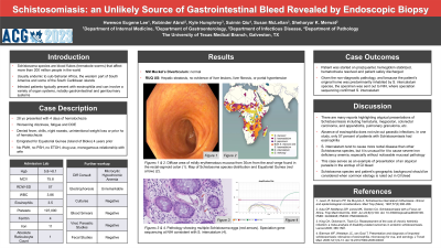
Case Description/Methods: A 28-year-old patient from Equatorial Guinea with no past medical history presented as a transfer from outside hospital with hemoglobin of 5.8 g/dL. The patient had been having severe fatigue for the past year, and significant hematochezia for the past week. He had no contributory surgical, family or social history. The patient was tachycardic with conjunctival pallor, but had otherwise unremarkable physical exam. Admission labs revealed iron deficiency anemia with hemoglobin of 8.1 g/dL. Other complete cell counts, coagulation studies, hemolysis studies, blood smear, fecal ova and parasites, and infectious workup were unremarkable. Imaging was unremarkable. Colonoscopy showed a diffuse area of mildly erythematous mucosa in the recto-sigmoid colon. Biopsy revealed colonic mucosa with calcified parasitic eggs, later identified to be Schistosoma intercalatum. The patient was subsequently treated with praziquantel with improvement of the symptoms and stabilization of hemoglobin. Patient was safely discharged with plans for follow up.
Discussion: There are reports highlighting atypical presentations of Schistosoma mansoni and hematobium species. Schistosoma intercalatum, though less prevalent, have been reported to cause rectal bleeding with inflammation of the rectum and colon. However, it is unusual for the parasite to cause such profound iron deficiency anemia as seen in our patient, especially without noticeable mucosal pathology.
Diagnosis of Schistosoma species is made on the presence of ova in feces or in biopsies of rectal mucosa. The endoscopic appearance of lesions is variable, ranging from granulomas to polyps to ulcerations. In non-endemic areas, it is often misdiagnosed as ulcerative colitis, Crohn’s disease, and ischemic colitis. Therefore, it is important to maintain low threshold for endoscopic biopsy in patients at risk for parasitic infection, including Schistosoma species.
In conclusion, this case serves as an example of presentation of an atypical parasite in the workup of GI bleed. Schistosoma species and a patient's geographic background should be considered when common etiology is ruled out in GI bleed.

Disclosures:
Hwewon E. Lee, DO1, Robinder Abrol, DO1, Suimin Qiu, MD, PhD2, Sheharyar Merwat, MBBS1, Kyle Humphrey, MD1. P0648 - Schistosomiasis: An Unlikely Source of Gastrointestinal Bleed Revealed by Endoscopic Biopsy, ACG 2023 Annual Scientific Meeting Abstracts. Vancouver, BC, Canada: American College of Gastroenterology.
1University of Texas Medical Branch, Galveston, TX; 2UTMB, Galveston, TX
Introduction: Although uncommon in the United States, Schistosoma species are blood flukes that affect more than 200 million in many developing countries including Africa. It is often misdiagnosed by physicians in non-endemic areas. Schistosomiasis can involve a variety of organ systems, notably gastrointestinal and genitourinary systems.
Case Description/Methods: A 28-year-old patient from Equatorial Guinea with no past medical history presented as a transfer from outside hospital with hemoglobin of 5.8 g/dL. The patient had been having severe fatigue for the past year, and significant hematochezia for the past week. He had no contributory surgical, family or social history. The patient was tachycardic with conjunctival pallor, but had otherwise unremarkable physical exam. Admission labs revealed iron deficiency anemia with hemoglobin of 8.1 g/dL. Other complete cell counts, coagulation studies, hemolysis studies, blood smear, fecal ova and parasites, and infectious workup were unremarkable. Imaging was unremarkable. Colonoscopy showed a diffuse area of mildly erythematous mucosa in the recto-sigmoid colon. Biopsy revealed colonic mucosa with calcified parasitic eggs, later identified to be Schistosoma intercalatum. The patient was subsequently treated with praziquantel with improvement of the symptoms and stabilization of hemoglobin. Patient was safely discharged with plans for follow up.
Discussion: There are reports highlighting atypical presentations of Schistosoma mansoni and hematobium species. Schistosoma intercalatum, though less prevalent, have been reported to cause rectal bleeding with inflammation of the rectum and colon. However, it is unusual for the parasite to cause such profound iron deficiency anemia as seen in our patient, especially without noticeable mucosal pathology.
Diagnosis of Schistosoma species is made on the presence of ova in feces or in biopsies of rectal mucosa. The endoscopic appearance of lesions is variable, ranging from granulomas to polyps to ulcerations. In non-endemic areas, it is often misdiagnosed as ulcerative colitis, Crohn’s disease, and ischemic colitis. Therefore, it is important to maintain low threshold for endoscopic biopsy in patients at risk for parasitic infection, including Schistosoma species.
In conclusion, this case serves as an example of presentation of an atypical parasite in the workup of GI bleed. Schistosoma species and a patient's geographic background should be considered when common etiology is ruled out in GI bleed.

Figure: A: Diffuse area of mildly erythematous mucosa from 30cm from the anal verge found in the rectal-sigmoid colon.
B: Map of Schistosoma species distribution and Equatorial Guinea (red arrow).
C: Pathology showing multiple Schistosoma eggs (red arrows).
D: Speciation gene sequencing at NIH consistent with S. intercalatum.
B: Map of Schistosoma species distribution and Equatorial Guinea (red arrow).
C: Pathology showing multiple Schistosoma eggs (red arrows).
D: Speciation gene sequencing at NIH consistent with S. intercalatum.
Disclosures:
Hwewon Lee indicated no relevant financial relationships.
Robinder Abrol indicated no relevant financial relationships.
Suimin Qiu indicated no relevant financial relationships.
Sheharyar Merwat indicated no relevant financial relationships.
Kyle Humphrey indicated no relevant financial relationships.
Hwewon E. Lee, DO1, Robinder Abrol, DO1, Suimin Qiu, MD, PhD2, Sheharyar Merwat, MBBS1, Kyle Humphrey, MD1. P0648 - Schistosomiasis: An Unlikely Source of Gastrointestinal Bleed Revealed by Endoscopic Biopsy, ACG 2023 Annual Scientific Meeting Abstracts. Vancouver, BC, Canada: American College of Gastroenterology.

