Sunday Poster Session
Category: GI Bleeding
P0650 - A Rare Cause of Upper GI Bleeding: Ampullary Dieulafoy Lesion
Sunday, October 22, 2023
3:30 PM - 7:00 PM PT
Location: Exhibit Hall

Has Audio

Annie Shergill, MD
Larkin Community Hospital
Miami, FL
Presenting Author(s)
Award: Presidential Poster Award
Annie Shergill, MD1, Abhishek Gulati, MD2
1Larkin Community Hospital, Miami, FL; 2Clovis Community Medical Center, Clovis, CA
Introduction: Ampullary Dieulafoy lesions (ADL) are an extremely rare cause of upper GI bleeding. ADLs can be very difficult to diagnose as they cannot be evaluated adequately with upper endoscopy (with forward viewing scope) or imaging modalities such as bleeding scans & angiography. We present a rare case of refractory upper GI bleeding secondary to an ADL.
Case Description/Methods: A 45-year old female with history of alcohol use presented with hematemesis, acute blood loss anemia with hemoglobin drop to 6 g/dL from her baseline of 12 g/dL. Initial upper endoscopy showed erosions in the gastric antrum, fresh blood in duodenum with no clear bleeding source. Patient continued to have hematemesis & underwent a bleeding scan which showed active bleeding in second portion of the duodenum. Patient was taken to Interventional Radiology suite for embolization however no active extravasation was noted on evaluation of Celiac and Superior Mesenteric Artery territory with an angiogram. Patient continued to bleed so a repeat upper endoscopy showed fresh blood in second portion of duodenum with brisk re-accumulation despite aggressive suctioning indicating active bleeding. Forward-viewing scope was switched to a side-viewing scope which revealed a spurting arterial bleed from ampulla with no underlying ulcer consistent with ADL (Figure 1). Hemostasis achieved with bipolar coagulation using gold probe.
Discussion: Extra-gastric dieulafoy lesions (DL) are most commonly found in the duodenum & account for 14-18% of cases. These lesions are mostly located in the duodenal bulb & are very rarely found in the second portion of the duodenum. Underlying pathophysiology of DL involves abnormality of anatomical arteriole under the mucosal surface which ruptures as the mucosa erodes, preceded by an insult such as alcohol or NSAID use. Extra-gastric DLs in the second portion of the duodenum pose a complex diagnostic challenge given the obscure location and sub-optimal evaluation of the periampullary area with a forward viewing scope. This necessitates evaluation with a side-viewing scope in order to clearly visualize the active area of bleeding & use therapeutic modalities such as bipolar coagulation, epinephrine injection and hemostatic clips. Successful endoscopic treatment of ADLs with a side-viewing scope has replaced the need for surgical intervention in these cases. Our case emphasizes on the importance of prompt evaluation with a side-viewing scope in case of refractory upper bleeding with a suspicious small bowel source.

Disclosures:
Annie Shergill, MD1, Abhishek Gulati, MD2. P0650 - A Rare Cause of Upper GI Bleeding: Ampullary Dieulafoy Lesion, ACG 2023 Annual Scientific Meeting Abstracts. Vancouver, BC, Canada: American College of Gastroenterology.
Annie Shergill, MD1, Abhishek Gulati, MD2
1Larkin Community Hospital, Miami, FL; 2Clovis Community Medical Center, Clovis, CA
Introduction: Ampullary Dieulafoy lesions (ADL) are an extremely rare cause of upper GI bleeding. ADLs can be very difficult to diagnose as they cannot be evaluated adequately with upper endoscopy (with forward viewing scope) or imaging modalities such as bleeding scans & angiography. We present a rare case of refractory upper GI bleeding secondary to an ADL.
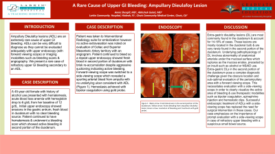
Case Description/Methods: A 45-year old female with history of alcohol use presented with hematemesis, acute blood loss anemia with hemoglobin drop to 6 g/dL from her baseline of 12 g/dL. Initial upper endoscopy showed erosions in the gastric antrum, fresh blood in duodenum with no clear bleeding source. Patient continued to have hematemesis & underwent a bleeding scan which showed active bleeding in second portion of the duodenum. Patient was taken to Interventional Radiology suite for embolization however no active extravasation was noted on evaluation of Celiac and Superior Mesenteric Artery territory with an angiogram. Patient continued to bleed so a repeat upper endoscopy showed fresh blood in second portion of duodenum with brisk re-accumulation despite aggressive suctioning indicating active bleeding. Forward-viewing scope was switched to a side-viewing scope which revealed a spurting arterial bleed from ampulla with no underlying ulcer consistent with ADL (Figure 1). Hemostasis achieved with bipolar coagulation using gold probe.
Discussion: Extra-gastric dieulafoy lesions (DL) are most commonly found in the duodenum & account for 14-18% of cases. These lesions are mostly located in the duodenal bulb & are very rarely found in the second portion of the duodenum. Underlying pathophysiology of DL involves abnormality of anatomical arteriole under the mucosal surface which ruptures as the mucosa erodes, preceded by an insult such as alcohol or NSAID use. Extra-gastric DLs in the second portion of the duodenum pose a complex diagnostic challenge given the obscure location and sub-optimal evaluation of the periampullary area with a forward viewing scope. This necessitates evaluation with a side-viewing scope in order to clearly visualize the active area of bleeding & use therapeutic modalities such as bipolar coagulation, epinephrine injection and hemostatic clips. Successful endoscopic treatment of ADLs with a side-viewing scope has replaced the need for surgical intervention in these cases. Our case emphasizes on the importance of prompt evaluation with a side-viewing scope in case of refractory upper bleeding with a suspicious small bowel source.

Figure: Figure 1: Black arrow: Fresh blood seen in the second portion of the duodenum; Yellow arrow: Active bleeding from ampullary dieulafoy lesion; Green arrow: resolution of bleeding post treatment with gold probe.
Disclosures:
Annie Shergill indicated no relevant financial relationships.
Abhishek Gulati indicated no relevant financial relationships.
Annie Shergill, MD1, Abhishek Gulati, MD2. P0650 - A Rare Cause of Upper GI Bleeding: Ampullary Dieulafoy Lesion, ACG 2023 Annual Scientific Meeting Abstracts. Vancouver, BC, Canada: American College of Gastroenterology.

