Sunday Poster Session
Category: Interventional Endoscopy
P0869 - Endoscopic Treatment of “Rabbit Ear” Pouch
Sunday, October 22, 2023
3:30 PM - 7:00 PM PT
Location: Exhibit Hall

Has Audio
- AF
Alisa Farokhian, MD
Columbia University
New York, NY
Presenting Author(s)
Award: Presidential Poster Award
Alisa Farokhian, MD1, Bo Shen, MD, FACG2
1Columbia University, New York, NY; 2Columbia University Medical Center, New York, NY
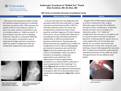
Introduction: Ileal pouch anal-anastomosis (IPAA) remains the standard surgical treatment for medically refractory ulcerative colitis. IPAA improves the quality of life for most patients, however it does not remain without complications such as a residual septum or “rabbit ear pouch”. A pouch septum may also result in bleeding, tenesmus, anal pain, or evacuation difficulty. In this case report we describe a case of apical pouch bridge syndrome caused by a rabbit ear septum configuration that was treated with septectomy endoscopically.
Case Description/Methods: A 53-year-old male who was diagnosed with ulcerative colitis (UC) and underwent a 2-stage restorative proctocolectomy IPAA in 2001 for medically refractory colitis. His course was complicated by anastomotic leak, chronic pouchitis, and later diagnosis of Crohn’s disease of the pouch. He was treated with infliximab, 6-mercaptopurine then ultimately switched to vedoluzimab. In 2020 he presented with dyschezia and tenesmus. Pouchoscopy was performed which was significant for a rabbit ear configuration of 10 cm. Barium defecography and anorectal manometry were performed (Figure 1) and the patient was sent for 10 sessions of biofeedback. With only 10% of resolution of symptoms, endoscopic augmentation was performed. First septecotomyty for pouch augmentation completed 3 cm, and the patient underwent a second septectomy for the remaining 7 cm along (figure 3) with another 10 sessions of biofeedback. There was significant improvement of symptoms after the second procedure and a repeat barium defecography (Figure 2) is shown below.
Discussion: Roughly 20% of IPAA patients experience structural complications after surgery. Although surgery has been the traditional therapy for these complications, endoscopic therapy has been seen to emerge as an alternative option. This “rabbit ear” configuration that was seen in our patient, can be caused by septum formation leading to dyschezia. Endoscopically the septum was cut using needle knife and 10 endoclips were placed. For our patient, this process was repeated three months after using the same technique. Barium defecography that was done after 3 septectomy treatments endoscopically showed improvement of the rabbit ear pouch with good transit into the afferent small bowel.

Disclosures:
Alisa Farokhian, MD1, Bo Shen, MD, FACG2. P0869 - Endoscopic Treatment of “Rabbit Ear” Pouch, ACG 2023 Annual Scientific Meeting Abstracts. Vancouver, BC, Canada: American College of Gastroenterology.
Alisa Farokhian, MD1, Bo Shen, MD, FACG2
1Columbia University, New York, NY; 2Columbia University Medical Center, New York, NY
Introduction: Ileal pouch anal-anastomosis (IPAA) remains the standard surgical treatment for medically refractory ulcerative colitis. IPAA improves the quality of life for most patients, however it does not remain without complications such as a residual septum or “rabbit ear pouch”. A pouch septum may also result in bleeding, tenesmus, anal pain, or evacuation difficulty. In this case report we describe a case of apical pouch bridge syndrome caused by a rabbit ear septum configuration that was treated with septectomy endoscopically.
Case Description/Methods: A 53-year-old male who was diagnosed with ulcerative colitis (UC) and underwent a 2-stage restorative proctocolectomy IPAA in 2001 for medically refractory colitis. His course was complicated by anastomotic leak, chronic pouchitis, and later diagnosis of Crohn’s disease of the pouch. He was treated with infliximab, 6-mercaptopurine then ultimately switched to vedoluzimab. In 2020 he presented with dyschezia and tenesmus. Pouchoscopy was performed which was significant for a rabbit ear configuration of 10 cm. Barium defecography and anorectal manometry were performed (Figure 1) and the patient was sent for 10 sessions of biofeedback. With only 10% of resolution of symptoms, endoscopic augmentation was performed. First septecotomyty for pouch augmentation completed 3 cm, and the patient underwent a second septectomy for the remaining 7 cm along (figure 3) with another 10 sessions of biofeedback. There was significant improvement of symptoms after the second procedure and a repeat barium defecography (Figure 2) is shown below.
Discussion: Roughly 20% of IPAA patients experience structural complications after surgery. Although surgery has been the traditional therapy for these complications, endoscopic therapy has been seen to emerge as an alternative option. This “rabbit ear” configuration that was seen in our patient, can be caused by septum formation leading to dyschezia. Endoscopically the septum was cut using needle knife and 10 endoclips were placed. For our patient, this process was repeated three months after using the same technique. Barium defecography that was done after 3 septectomy treatments endoscopically showed improvement of the rabbit ear pouch with good transit into the afferent small bowel.

Figure: Figures associated with Rabbit ear
Disclosures:
Alisa Farokhian indicated no relevant financial relationships.
Bo Shen: AbbVie – Consultant. Janssen – Consultant. Takeda – Consultant.
Alisa Farokhian, MD1, Bo Shen, MD, FACG2. P0869 - Endoscopic Treatment of “Rabbit Ear” Pouch, ACG 2023 Annual Scientific Meeting Abstracts. Vancouver, BC, Canada: American College of Gastroenterology.

