Sunday Poster Session
Category: Interventional Endoscopy
P0872 - First Reported Case of Endoscopic En Bloc Resection of Esophageal Liposarcoma
Sunday, October 22, 2023
3:30 PM - 7:00 PM PT
Location: Exhibit Hall

Has Audio

Taher Jamali, MD
Henry Ford Health
Farmington Hills, MI
Presenting Author(s)
Award: Presidential Poster Award
Taher Jamali, MD1, Jason Pimentel, MD2, Kyle Perry, MD2, Jack Tocco, MD3, Sheela Tejwani, MD2, Ross Mayerhoff, MD2, Robert Pompa, MD4
1Henry Ford Health, Farmington Hills, MI; 2Henry Ford Health, Detroit, MI; 3Pinnacle GI Partners, Troy, MI; 4Henry Ford Hospital, Detroit, MI
Introduction: Well-differentiated sarcomas of the esophagus have been rarely reported in the literature with less than 100 cases reported to date. Here, we present the first described case of esophageal liposarcoma removed via en-bloc endoscopic mucosal resection (EMR).
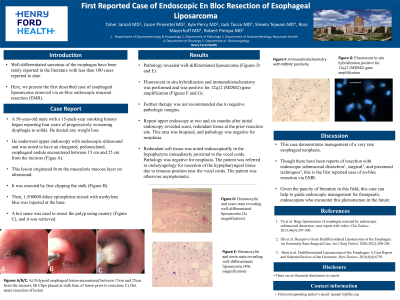
Case Description/Methods: A 58-year-old male with a 15-pack-year smoking history began reported four years of progressively worsening dysphagia to solids. He denied any weight loss. He underwent upper endoscopy with endoscopic ultrasound and was noted to have an elongated, pedunculated, esophageal nodule encountered between 15cm and 25cm from the incisors. This lesion originated from the muscularis mucosa layer on ultrasound. It was resected by first clipping the stalk. Then, 1:500000 dilute epinephrine mixed with methylene blue was injected at the base. A hot snare was used to resect the polyp using cautery, and it was retrieved. Pathology revealed well-differentiated liposarcoma. Fluorescent in situ hybridization was performed and was positive for 12q12 (MDM2) gene amplification. Further therapy was not recommended due to negative pathologic margins. Repeat upper endoscopy at two and six months after initial endoscopy revealed scant, redundant tissue at the prior resection site. This area was biopsied, and pathology was negative for neoplasia. Redundant soft tissue was noted endoscopically in the hypopharynx immediately proximal to the vocal cords. Pathology was negative for neoplasia. The patient was referred to otolaryngology for resection of the hypopharyngeal tissue due to tenuous position near the vocal cords. The patient was otherwise asymptomatic.
Discussion: This case demonstrates management of a very rare esophageal neoplasia. Though there have been reports of resection with endoscopic submucosal dissection1, surgical2, and piecemeal techniques3, this is the first reported case of en-bloc resection via EMR. Given the paucity of literature in this field, this case can help to guide endoscopic management for therapeutic endoscopists who encounter this phenomenon in the future.
1. Yo et al. Huge liposarcoma of esophagus resected by endoscopic submucosal dissection: case report with video. Clin Endosc. 2013;46(3):297-300.
2. Shi et al. Deceptive Giant Dedifferentiated Liposarcoma of the Esophagus: An Extremely Rare Surgical Case. Int J Surg Pathol. 2020;28(2):200-205.
3. Brett et al. Dedifferentiated Liposarcoma of the Esophagus: A Case Report and Selected Review of the Literature. Rare Tumors. 2016;8(4):6791.

Disclosures:
Taher Jamali, MD1, Jason Pimentel, MD2, Kyle Perry, MD2, Jack Tocco, MD3, Sheela Tejwani, MD2, Ross Mayerhoff, MD2, Robert Pompa, MD4. P0872 - First Reported Case of Endoscopic En Bloc Resection of Esophageal Liposarcoma, ACG 2023 Annual Scientific Meeting Abstracts. Vancouver, BC, Canada: American College of Gastroenterology.
Taher Jamali, MD1, Jason Pimentel, MD2, Kyle Perry, MD2, Jack Tocco, MD3, Sheela Tejwani, MD2, Ross Mayerhoff, MD2, Robert Pompa, MD4
1Henry Ford Health, Farmington Hills, MI; 2Henry Ford Health, Detroit, MI; 3Pinnacle GI Partners, Troy, MI; 4Henry Ford Hospital, Detroit, MI
Introduction: Well-differentiated sarcomas of the esophagus have been rarely reported in the literature with less than 100 cases reported to date. Here, we present the first described case of esophageal liposarcoma removed via en-bloc endoscopic mucosal resection (EMR).
Case Description/Methods: A 58-year-old male with a 15-pack-year smoking history began reported four years of progressively worsening dysphagia to solids. He denied any weight loss. He underwent upper endoscopy with endoscopic ultrasound and was noted to have an elongated, pedunculated, esophageal nodule encountered between 15cm and 25cm from the incisors. This lesion originated from the muscularis mucosa layer on ultrasound. It was resected by first clipping the stalk. Then, 1:500000 dilute epinephrine mixed with methylene blue was injected at the base. A hot snare was used to resect the polyp using cautery, and it was retrieved. Pathology revealed well-differentiated liposarcoma. Fluorescent in situ hybridization was performed and was positive for 12q12 (MDM2) gene amplification. Further therapy was not recommended due to negative pathologic margins. Repeat upper endoscopy at two and six months after initial endoscopy revealed scant, redundant tissue at the prior resection site. This area was biopsied, and pathology was negative for neoplasia. Redundant soft tissue was noted endoscopically in the hypopharynx immediately proximal to the vocal cords. Pathology was negative for neoplasia. The patient was referred to otolaryngology for resection of the hypopharyngeal tissue due to tenuous position near the vocal cords. The patient was otherwise asymptomatic.
Discussion: This case demonstrates management of a very rare esophageal neoplasia. Though there have been reports of resection with endoscopic submucosal dissection1, surgical2, and piecemeal techniques3, this is the first reported case of en-bloc resection via EMR. Given the paucity of literature in this field, this case can help to guide endoscopic management for therapeutic endoscopists who encounter this phenomenon in the future.
1. Yo et al. Huge liposarcoma of esophagus resected by endoscopic submucosal dissection: case report with video. Clin Endosc. 2013;46(3):297-300.
2. Shi et al. Deceptive Giant Dedifferentiated Liposarcoma of the Esophagus: An Extremely Rare Surgical Case. Int J Surg Pathol. 2020;28(2):200-205.
3. Brett et al. Dedifferentiated Liposarcoma of the Esophagus: A Case Report and Selected Review of the Literature. Rare Tumors. 2016;8(4):6791.

Figure: Figure 1: A) Polypoid esophageal lesion encountered between 15cm and 25cm from the incisors; B) Clips placed at stalk base of lesion prior to resection; C) Hot snare resection of lesion; D) Fluorescent in situ hybridization positive for 12q12 (MDM2) gene amplification
Disclosures:
Taher Jamali indicated no relevant financial relationships.
Jason Pimentel indicated no relevant financial relationships.
Kyle Perry indicated no relevant financial relationships.
Jack Tocco indicated no relevant financial relationships.
Sheela Tejwani indicated no relevant financial relationships.
Ross Mayerhoff indicated no relevant financial relationships.
Robert Pompa indicated no relevant financial relationships.
Taher Jamali, MD1, Jason Pimentel, MD2, Kyle Perry, MD2, Jack Tocco, MD3, Sheela Tejwani, MD2, Ross Mayerhoff, MD2, Robert Pompa, MD4. P0872 - First Reported Case of Endoscopic En Bloc Resection of Esophageal Liposarcoma, ACG 2023 Annual Scientific Meeting Abstracts. Vancouver, BC, Canada: American College of Gastroenterology.


