Sunday Poster Session
Category: Interventional Endoscopy
P0873 - A Rare Case of Esophageal Obstruction After Rupture of an Esophageal Duplication Cyst
Sunday, October 22, 2023
3:30 PM - 7:00 PM PT
Location: Exhibit Hall

Has Audio

Taher Jamali, MD
Henry Ford Health
Farmington Hills, MI
Presenting Author(s)
Award: Presidential Poster Award
Taher Jamali, MD1, Miguel Alvelo-Rivera, MD2, Mazen Elatrache, MD3
1Henry Ford Health, Farmington Hills, MI; 2Henry Ford Health, Detroit, MI; 3Henry Ford Hospital, Detroit, MI
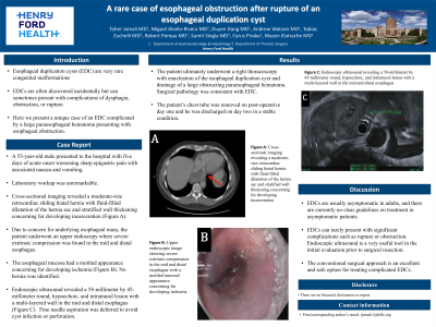
Introduction: Esophageal duplication cysts (EDC) are very rare congenital malformations, which often are discovered incidentally but can sometimes present with complications of dysphagia, obstruction, or rupture. Here we present a unique case of an EDC complicated by a large paraesophageal hematoma presenting with esophageal obstruction.
Case Description/Methods: A 55-year-old male presented to the hospital with five days of acute onset worsening sharp epigastric pain with associated nausea and vomiting. Laboratory workup was unremarkable. Cross-sectional imaging revealed a moderate-size retrocardiac sliding hiatal hernia with fluid-filled dilatation of the hernia sac and stratified wall thickening concerning for developing incarceration (Figure 1A). Due to concern for underlying esophageal mass, the patient underwent an upper endoscopy where severe extrinsic compression was found in the mid and distal esophagus. The esophageal mucosa had a mottled appearance concerning for developing ischemia (Figure 1B). No hernia was identified. Endoscopic ultrasound revealed a 58-millimeter by 45-millimeter round, hypoechoic, and intramural lesion with a multi-layered wall in the mid and distal esophagus (Figure 1C). Fine needle aspiration was deferred to avoid cyst infection or perforation. The patient ultimately underwent a right thoracoscopy with enucleation of the esophageal duplication cyst and drainage of a large obstructing paraesophageal hematoma. Surgical pathology was consistent with EDC. The patient’s chest tube was removed on post-operative day one and he was discharged on day two in a stable condition.
Discussion: EDCs are usually asymptomatic in adults, and there are currently no clear guidelines on treatment in asymptomatic patients. EDCs can rarely present with significant complications such as rupture or obstruction. Endoscopic ultrasound is a very useful tool in the initial evaluation prior to surgical resection. The conventional surgical approach is an excellent and safe option for treating complicated EDCs.

Disclosures:
Taher Jamali, MD1, Miguel Alvelo-Rivera, MD2, Mazen Elatrache, MD3. P0873 - A Rare Case of Esophageal Obstruction After Rupture of an Esophageal Duplication Cyst, ACG 2023 Annual Scientific Meeting Abstracts. Vancouver, BC, Canada: American College of Gastroenterology.
Taher Jamali, MD1, Miguel Alvelo-Rivera, MD2, Mazen Elatrache, MD3
1Henry Ford Health, Farmington Hills, MI; 2Henry Ford Health, Detroit, MI; 3Henry Ford Hospital, Detroit, MI
Introduction: Esophageal duplication cysts (EDC) are very rare congenital malformations, which often are discovered incidentally but can sometimes present with complications of dysphagia, obstruction, or rupture. Here we present a unique case of an EDC complicated by a large paraesophageal hematoma presenting with esophageal obstruction.
Case Description/Methods: A 55-year-old male presented to the hospital with five days of acute onset worsening sharp epigastric pain with associated nausea and vomiting. Laboratory workup was unremarkable. Cross-sectional imaging revealed a moderate-size retrocardiac sliding hiatal hernia with fluid-filled dilatation of the hernia sac and stratified wall thickening concerning for developing incarceration (Figure 1A). Due to concern for underlying esophageal mass, the patient underwent an upper endoscopy where severe extrinsic compression was found in the mid and distal esophagus. The esophageal mucosa had a mottled appearance concerning for developing ischemia (Figure 1B). No hernia was identified. Endoscopic ultrasound revealed a 58-millimeter by 45-millimeter round, hypoechoic, and intramural lesion with a multi-layered wall in the mid and distal esophagus (Figure 1C). Fine needle aspiration was deferred to avoid cyst infection or perforation. The patient ultimately underwent a right thoracoscopy with enucleation of the esophageal duplication cyst and drainage of a large obstructing paraesophageal hematoma. Surgical pathology was consistent with EDC. The patient’s chest tube was removed on post-operative day one and he was discharged on day two in a stable condition.
Discussion: EDCs are usually asymptomatic in adults, and there are currently no clear guidelines on treatment in asymptomatic patients. EDCs can rarely present with significant complications such as rupture or obstruction. Endoscopic ultrasound is a very useful tool in the initial evaluation prior to surgical resection. The conventional surgical approach is an excellent and safe option for treating complicated EDCs.

Figure: Figure 1: A) Cross-sectional imaging revealing a moderate-size retrocardiac sliding hiatal hernia with fluid-filled dilatation of the hernia sac and stratified wall thickening concerning for developing incarceration; B) Upper endoscopic image showing severe extrinsic compression in the mid and distal esophagus with a mottled mucosal appearance concerning for developing ischemia; C) Endoscopic ultrasound revealing a 58-millimeter by 45-millimeter round, hypoechoic, and intramural lesion with a multi-layered wall in the mid and distal esophagus
Disclosures:
Taher Jamali indicated no relevant financial relationships.
Miguel Alvelo-Rivera indicated no relevant financial relationships.
Mazen Elatrache indicated no relevant financial relationships.
Taher Jamali, MD1, Miguel Alvelo-Rivera, MD2, Mazen Elatrache, MD3. P0873 - A Rare Case of Esophageal Obstruction After Rupture of an Esophageal Duplication Cyst, ACG 2023 Annual Scientific Meeting Abstracts. Vancouver, BC, Canada: American College of Gastroenterology.

