Sunday Poster Session
Category: Interventional Endoscopy
P0892 - Endoscopic Ultrasound Guided Subhepatic Biloma Drainage in a Patient With Partial Hepatectomy With Prior Roux-En-Y Anatomy
Sunday, October 22, 2023
3:30 PM - 7:00 PM PT
Location: Exhibit Hall

Has Audio
- RB
Ramanpreet Bajwa, DO
Cleveland Clinic Foundation
Shaker Heights, Ohio
Presenting Author(s)
Ramanpreet Bajwa, DO, Lovepreet Singh, MD, Prabhleen Chahal, MD
Cleveland Clinic Foundation, Cleveland, OH
Introduction: Biloma formation following liver surgery is a major complication associated with great morbidity and mortality. Ultrasound or computed tomography (CT) guided drainage is the preferred modality. We present a case of a symptomatic subhepatic biloma in a patient with recent partial hepatectomy with altered anatomy due to previous Roux-en-Y surgery.
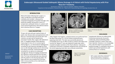
Case Description/Methods: 70 year old male with past medical history of peptic ulcer disease status post gastrectomy with Roux-en-Y reconstruction and recent liver mass resection via open partial hepatectomy presented 4 weeks after surgery with abdominal pain. CT imaging revealed a fluid collection measuring 6.5 x 6.6 x 9.5 cm concerning for biloma (Figure 1). Interventional radiology was consulted for percutaneous drainage but there was no safe drainage window due to intervening vasculature and bowel. Gastroenterology was consulted for an opinion on endoscopic management. Review of imaging revealed that the collection could be drained from the Roux limb. The linear echoendoscope was advanced with the patient on the fluoroscopy table. The large collection was localized from the Roux limb in close proximity to the small bowel in a subhepatic location. After doppler interrogation, cystojejunostomy was performed from the Roux limb using a 15 x 10 mm cautery enhanced lumen apposing metal stent. The stent was successfully placed and immediately there was an efflux of bilious contents with debris and necrotic fat. Repeat CT scan was obtained 4 weeks later which revealed complete resolution of the fluid collection (Figure 1). Subsequent endoscopy confirmed a completely collapsed cavity and removal of the lumen apposing metal stent was achieved uneventfully.
Discussion: Symptomatic bilomas are preferentially drained percutaneously. Successful endoscopic ultrasound (EUS) guided drainage of bilomas in select cases have been reported. However to our knowledge, this is the first report of successful EUS guided drainage of a subhepatic biloma in a patient post partial hepatectomy with previous Roux-en-Y anatomy.

Disclosures:
Ramanpreet Bajwa, DO, Lovepreet Singh, MD, Prabhleen Chahal, MD. P0892 - Endoscopic Ultrasound Guided Subhepatic Biloma Drainage in a Patient With Partial Hepatectomy With Prior Roux-En-Y Anatomy, ACG 2023 Annual Scientific Meeting Abstracts. Vancouver, BC, Canada: American College of Gastroenterology.
Cleveland Clinic Foundation, Cleveland, OH
Introduction: Biloma formation following liver surgery is a major complication associated with great morbidity and mortality. Ultrasound or computed tomography (CT) guided drainage is the preferred modality. We present a case of a symptomatic subhepatic biloma in a patient with recent partial hepatectomy with altered anatomy due to previous Roux-en-Y surgery.
Case Description/Methods: 70 year old male with past medical history of peptic ulcer disease status post gastrectomy with Roux-en-Y reconstruction and recent liver mass resection via open partial hepatectomy presented 4 weeks after surgery with abdominal pain. CT imaging revealed a fluid collection measuring 6.5 x 6.6 x 9.5 cm concerning for biloma (Figure 1). Interventional radiology was consulted for percutaneous drainage but there was no safe drainage window due to intervening vasculature and bowel. Gastroenterology was consulted for an opinion on endoscopic management. Review of imaging revealed that the collection could be drained from the Roux limb. The linear echoendoscope was advanced with the patient on the fluoroscopy table. The large collection was localized from the Roux limb in close proximity to the small bowel in a subhepatic location. After doppler interrogation, cystojejunostomy was performed from the Roux limb using a 15 x 10 mm cautery enhanced lumen apposing metal stent. The stent was successfully placed and immediately there was an efflux of bilious contents with debris and necrotic fat. Repeat CT scan was obtained 4 weeks later which revealed complete resolution of the fluid collection (Figure 1). Subsequent endoscopy confirmed a completely collapsed cavity and removal of the lumen apposing metal stent was achieved uneventfully.
Discussion: Symptomatic bilomas are preferentially drained percutaneously. Successful endoscopic ultrasound (EUS) guided drainage of bilomas in select cases have been reported. However to our knowledge, this is the first report of successful EUS guided drainage of a subhepatic biloma in a patient post partial hepatectomy with previous Roux-en-Y anatomy.

Figure: Figure 1. Subhepatic Biloma Prior to Drainage (A) followed by Resolution of Biloma after EUS guided Drainage (B)
Disclosures:
Ramanpreet Bajwa indicated no relevant financial relationships.
Lovepreet Singh indicated no relevant financial relationships.
Prabhleen Chahal indicated no relevant financial relationships.
Ramanpreet Bajwa, DO, Lovepreet Singh, MD, Prabhleen Chahal, MD. P0892 - Endoscopic Ultrasound Guided Subhepatic Biloma Drainage in a Patient With Partial Hepatectomy With Prior Roux-En-Y Anatomy, ACG 2023 Annual Scientific Meeting Abstracts. Vancouver, BC, Canada: American College of Gastroenterology.
