Sunday Poster Session
Category: Interventional Endoscopy
P0895 - Abdominal Ganglioneuroma Diagnosed With Fine Needle Biopsy on Endoscopic Ultrasound
Sunday, October 22, 2023
3:30 PM - 7:00 PM PT
Location: Exhibit Hall

Has Audio

Sangini Tolia, MD
Trinity Health St. Joseph Mercy Hospital-Ann Arbor
Ann Arbor, MI
Presenting Author(s)
Sangini Tolia, MD1, Michael Zijlstra, DO1, Ali Yazdani, MD2, Mohannad Abousaleh, MD2
1Trinity Health St. Joseph Mercy Hospital-Ann Arbor, Ann Arbor, MI; 2Huron Gastroenterology, Ann Arbor, MI
Introduction: Ganglioneuromas are rare, benign tumors that originate from neural crest cells. Although most patients are asymptomatic, this tumor can cause vague symptoms such as pain, vomiting, and diarrhea. There are few cases reported of ganglioneuroma diagnosed with endoscopic ultrasound (EUS) and fine needle biopsy (FNB). Here, we present a patient experiencing back pain and diarrhea, who was found to have an abdominal ganglioneuroma.
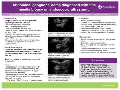
Case Description/Methods: A 70 year old female presented to her primary care physician with a 30 lb unintentional weight loss over the past year. She had watery stools 2-3 times per day, increased flatulence, and left sided thoracic back pain. She had no pertinent medical or family history, and serum laboratory workup was unremarkable. An abdominal computed tomography (CT) scan showed a 1 cm length of moderate severity narrowing of the superior mesenteric artery (SMA), with edema or soft tissue changes around the SMA extending into the mesentery. An abdominal MRI was also obtained, redemonstrating an ill-defined retroperitoneal soft tissue mass encasing the upper abdominal aorta, central celiac artery, and SMA. The differential included vasculitis and neoplastic infiltration but remained broad, prompting further imaging with positron emission tomography (PET) and Dotate PET CT scans. Both were negative for tracer uptake. The patient was then referred to gastroenterology. She underwent an EUS, revealing a 20 x 30 mm soft tissue mass encasing the celiac artery extending up to the splenic artery and involving the SMA. FNB was performed via a transgastric approach. Pathology results revealed a ganglioneuroma. The case was presented at tumor board, and the recommendation was made to monitor with serial imaging and refer to a sarcoma/soft tissue tumor clinic.
Discussion: Ganglioneuromas have a variable clinical presentation. They develop along the sympathetic chain in both pediatric and adult populations, and may present with vague symptoms. Interestingly, ganglioneuromas have been known to encase blood vessels without compromising the lumen. In this case, the patient was symptomatic and had evidence of mass effect. Given the lack of specific characteristics on imaging, histopathology analysis is necessary in order to make a diagnosis. This case is only one of few others that utilized EUS with FNB to identify ganglioneuroma. The procedure offers a unique opportunity to make the diagnosis in a less invasive approach, after which curative surgical options can be explored.

Disclosures:
Sangini Tolia, MD1, Michael Zijlstra, DO1, Ali Yazdani, MD2, Mohannad Abousaleh, MD2. P0895 - Abdominal Ganglioneuroma Diagnosed With Fine Needle Biopsy on Endoscopic Ultrasound, ACG 2023 Annual Scientific Meeting Abstracts. Vancouver, BC, Canada: American College of Gastroenterology.
1Trinity Health St. Joseph Mercy Hospital-Ann Arbor, Ann Arbor, MI; 2Huron Gastroenterology, Ann Arbor, MI
Introduction: Ganglioneuromas are rare, benign tumors that originate from neural crest cells. Although most patients are asymptomatic, this tumor can cause vague symptoms such as pain, vomiting, and diarrhea. There are few cases reported of ganglioneuroma diagnosed with endoscopic ultrasound (EUS) and fine needle biopsy (FNB). Here, we present a patient experiencing back pain and diarrhea, who was found to have an abdominal ganglioneuroma.
Case Description/Methods: A 70 year old female presented to her primary care physician with a 30 lb unintentional weight loss over the past year. She had watery stools 2-3 times per day, increased flatulence, and left sided thoracic back pain. She had no pertinent medical or family history, and serum laboratory workup was unremarkable. An abdominal computed tomography (CT) scan showed a 1 cm length of moderate severity narrowing of the superior mesenteric artery (SMA), with edema or soft tissue changes around the SMA extending into the mesentery. An abdominal MRI was also obtained, redemonstrating an ill-defined retroperitoneal soft tissue mass encasing the upper abdominal aorta, central celiac artery, and SMA. The differential included vasculitis and neoplastic infiltration but remained broad, prompting further imaging with positron emission tomography (PET) and Dotate PET CT scans. Both were negative for tracer uptake. The patient was then referred to gastroenterology. She underwent an EUS, revealing a 20 x 30 mm soft tissue mass encasing the celiac artery extending up to the splenic artery and involving the SMA. FNB was performed via a transgastric approach. Pathology results revealed a ganglioneuroma. The case was presented at tumor board, and the recommendation was made to monitor with serial imaging and refer to a sarcoma/soft tissue tumor clinic.
Discussion: Ganglioneuromas have a variable clinical presentation. They develop along the sympathetic chain in both pediatric and adult populations, and may present with vague symptoms. Interestingly, ganglioneuromas have been known to encase blood vessels without compromising the lumen. In this case, the patient was symptomatic and had evidence of mass effect. Given the lack of specific characteristics on imaging, histopathology analysis is necessary in order to make a diagnosis. This case is only one of few others that utilized EUS with FNB to identify ganglioneuroma. The procedure offers a unique opportunity to make the diagnosis in a less invasive approach, after which curative surgical options can be explored.

Figure: A. Fine needle biopsy of soft tissue thickening encasing the celiac artery
B. Fine needle biopsy of soft tissue thickening encasing the celiac and superior mesenteric arteries
C. EUS image with evidence of soft tissue mass encasing the celiac and superior mesenteric arteries near their take off from the abdominal aorta
B. Fine needle biopsy of soft tissue thickening encasing the celiac and superior mesenteric arteries
C. EUS image with evidence of soft tissue mass encasing the celiac and superior mesenteric arteries near their take off from the abdominal aorta
Disclosures:
Sangini Tolia indicated no relevant financial relationships.
Michael Zijlstra indicated no relevant financial relationships.
Ali Yazdani indicated no relevant financial relationships.
Mohannad Abousaleh indicated no relevant financial relationships.
Sangini Tolia, MD1, Michael Zijlstra, DO1, Ali Yazdani, MD2, Mohannad Abousaleh, MD2. P0895 - Abdominal Ganglioneuroma Diagnosed With Fine Needle Biopsy on Endoscopic Ultrasound, ACG 2023 Annual Scientific Meeting Abstracts. Vancouver, BC, Canada: American College of Gastroenterology.
