Sunday Poster Session
Category: Interventional Endoscopy
P0896 - Endoscopic Management of a Large Gastric Wall Defect Due to Anastomotic Marginal Ulcer Perforation
Sunday, October 22, 2023
3:30 PM - 7:00 PM PT
Location: Exhibit Hall

Has Audio

Bobby Jacob, MD
HCA Florida Largo Hospital
Largo, FL
Presenting Author(s)
Bobby Jacob, MD1, Shivani Trivedi, DO2, Vikas S. Sethi, DO3, Suhail Kayyali, DO4, Meir Mizrahi, MD4
1HCA Florida Largo Hospital, Largo, FL; 2Largo HCA Healthcare, Clearwater, FL; 3Largo Medical Center, Clearwater, FL; 4Largo Medical Center, Largo, FL
Introduction: Endoscopic management plays a crucial role in the treatment of anastomotic marginal gastric ulcer perforation, offering several benefits compared to traditional surgical interventions. The use of endoscopy allows for direct visualization of the ulcer site, facilitating accurate assessment of the perforation extent and characteristics. We describe a case involving a significant gastric wall defect resulting from a perforated anastomotic marginal ulcer, which was successfully managed using a distinctive endoscopic suturing technique.
Case Description/Methods: A 65-year-old Caucasian female with a complex medical history, including signet ring cell gastric cancer status post near total gastrectomy, and a recent sigmoid cancer diagnosis, underwent left hemicolectomy two weeks prior. The patient presented with fevers, and CT imaging revealed a pelvic abscess. An exploratory laparotomy confirmed an anastomotic leak, leading to partial colectomy, colostomy creation, and washout. The hospital course was further complicated by a perforated marginal gastroenteric anastomotic ulcer, resulting in a large defect in the anterior stomach. Following multidisciplinary consultation, the decision was made to explore endoscopic treatment options. Diagnostic endoscopy identified a large gastric wall defect near the gastrojejunostomy (GJ) anastomosis. Over the wire guided placement of an esophageal dilatation balloon across the GJ lumen was performed under endoscopic and fluoroscopic imaging. Subsequently, the balloon was inflated to 18mm to maintain patency of the GJ limb during endoscopic suturing. Two sutures were successfully placed to close the mucosal defect, and contrast injection confirmed satisfactory closure with no extravasation on fluoroscopy.
Discussion: Endoscopic interventions are valuable for both diagnosis and treatment of gastric perforation. This enables precise placement of clips or sutures to achieve effective closure, reducing the risk of persistent leakage and complications such as peritonitis or abscess formation. Adjunctive procedures like endoscopic vacuum therapy or stent insertion can be combined with endoscopic techniques to optimize outcomes in complex cases. However, large or complex perforations, extensive contamination, or necrosis may require surgical intervention and endoscopic management. Close collaboration between endoscopists and surgeons is crucial to determine the most suitable approach for each case.

Disclosures:
Bobby Jacob, MD1, Shivani Trivedi, DO2, Vikas S. Sethi, DO3, Suhail Kayyali, DO4, Meir Mizrahi, MD4. P0896 - Endoscopic Management of a Large Gastric Wall Defect Due to Anastomotic Marginal Ulcer Perforation, ACG 2023 Annual Scientific Meeting Abstracts. Vancouver, BC, Canada: American College of Gastroenterology.
1HCA Florida Largo Hospital, Largo, FL; 2Largo HCA Healthcare, Clearwater, FL; 3Largo Medical Center, Clearwater, FL; 4Largo Medical Center, Largo, FL
Introduction: Endoscopic management plays a crucial role in the treatment of anastomotic marginal gastric ulcer perforation, offering several benefits compared to traditional surgical interventions. The use of endoscopy allows for direct visualization of the ulcer site, facilitating accurate assessment of the perforation extent and characteristics. We describe a case involving a significant gastric wall defect resulting from a perforated anastomotic marginal ulcer, which was successfully managed using a distinctive endoscopic suturing technique.
Case Description/Methods: A 65-year-old Caucasian female with a complex medical history, including signet ring cell gastric cancer status post near total gastrectomy, and a recent sigmoid cancer diagnosis, underwent left hemicolectomy two weeks prior. The patient presented with fevers, and CT imaging revealed a pelvic abscess. An exploratory laparotomy confirmed an anastomotic leak, leading to partial colectomy, colostomy creation, and washout. The hospital course was further complicated by a perforated marginal gastroenteric anastomotic ulcer, resulting in a large defect in the anterior stomach. Following multidisciplinary consultation, the decision was made to explore endoscopic treatment options. Diagnostic endoscopy identified a large gastric wall defect near the gastrojejunostomy (GJ) anastomosis. Over the wire guided placement of an esophageal dilatation balloon across the GJ lumen was performed under endoscopic and fluoroscopic imaging. Subsequently, the balloon was inflated to 18mm to maintain patency of the GJ limb during endoscopic suturing. Two sutures were successfully placed to close the mucosal defect, and contrast injection confirmed satisfactory closure with no extravasation on fluoroscopy.
Discussion: Endoscopic interventions are valuable for both diagnosis and treatment of gastric perforation. This enables precise placement of clips or sutures to achieve effective closure, reducing the risk of persistent leakage and complications such as peritonitis or abscess formation. Adjunctive procedures like endoscopic vacuum therapy or stent insertion can be combined with endoscopic techniques to optimize outcomes in complex cases. However, large or complex perforations, extensive contamination, or necrosis may require surgical intervention and endoscopic management. Close collaboration between endoscopists and surgeons is crucial to determine the most suitable approach for each case.

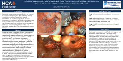
Figure: Image A: Gastric wall perforation adjacent to the anastomotic site. Image B: Endoscopic suturing of gastric wall defect with a dilated 18mm esophageal balloon to maintain patency and prevent inadvertent suturing of the alimentary limb. Image C and D: Status post endoscopic closure of the gastric wall defect.
Disclosures:
Bobby Jacob indicated no relevant financial relationships.
Shivani Trivedi indicated no relevant financial relationships.
Vikas Sethi indicated no relevant financial relationships.
Suhail Kayyali indicated no relevant financial relationships.
Meir Mizrahi: Boston Scientific – Consultant. COOK medical – Consultant. Fujifilm – Consultant. Medtronic – Consultant.
Bobby Jacob, MD1, Shivani Trivedi, DO2, Vikas S. Sethi, DO3, Suhail Kayyali, DO4, Meir Mizrahi, MD4. P0896 - Endoscopic Management of a Large Gastric Wall Defect Due to Anastomotic Marginal Ulcer Perforation, ACG 2023 Annual Scientific Meeting Abstracts. Vancouver, BC, Canada: American College of Gastroenterology.
