Sunday Poster Session
Category: Liver
P0999 - A Rare Case of Esophageal Hepatoid Adenocarcinoma With Lung and Liver Metastases Mimicking Hepatocellular Carcinoma
Sunday, October 22, 2023
3:30 PM - 7:00 PM PT
Location: Exhibit Hall

Has Audio

Maya Patel, MS, MD
Emory University School of Medicine
Atlanta, GA
Presenting Author(s)
Award: Presidential Poster Award
Maya Patel, MS, MD1, Victoria Earl, MD1, Sara Levy, MD2, Jigna Jani, MD2, Anand Shah, MD3
1Emory University School of Medicine, Atlanta, GA; 2Atlanta VA Medical Center, Decatur, GA; 3Emory University School of Medicine, Decatur, GA
Introduction: Hepatoid adenocarcinoma (HAC) is a rare, aggressive, extrahepatic tumor with hepatocellular differentiation that classically produces alpha-fetoprotein (AFP). These tumors are most commonly found in stomach but are unusual to arise from the esophagus. Early identification of HAC can be difficult given its similar morphology to hepatocellular carcinoma (HCC). Herein we present the case of primary esophageal HAC with metastases to the liver and lung initially concerning for primary HCC.
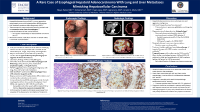
Case Description/Methods: A 68-year-old African American male with a medical history of cardiovascular disease, previous tobacco use, and without underlying chronic liver disease presented with 2 months of left-sided chest pain, nonproductive cough, early satiety, and unintentional weight loss. On physical examination, he had bibasilar pulmonary crackles. His abdomen had no appreciable hepatosplenomegaly nor did he have stigmata of chronic liver disease. Laboratory findings included an AFP level >51,000 ng/mL. Computed tomography angiography (CTA) of the chest demonstrated distal esophageal wall thickening with heterogenous enhancement, pulmonary, hilar, mediastinal lymphadenopathy, and hepatic lesions. Positron emission tomography-computed tomography (PET-CT) suggested primary esophageal malignancy with lung, liver, and lymph node metastases.
Esophagogastroduodenoscopy (EGD) was performed and a 7-centimenter distal esophageal mass extending to the gastric cardia was seen. Biopsy revealed carcinoma with hepatocellular differentiation and marked surface ulceration and necrosis. A fine needle biopsy of a liver lesion revealed slightly different morphology but the immunohistochemical (IHC) staining was similar for both sites: positive for glypican-3 and CDX2, and negative for CD-7, CD-20, and HepPar. These findings, coupled with the patient's lack of HCC risk factors, were ultimately favored to represent metastatic HAC. Unfortunately, the patient ultimately passed away from sequelae of his disease prior to initiation of treatment.
Discussion: Hepatoid adenocarcinoma is an aggressive tumor that is rarely found in the esophagus. Given its similar morphology to hepatocellular carcinoma and its association with elevated AFP levels, it can be difficult to differentiate the two malignancies. Diagnosis is possible with careful histopathologic examination and IHC staining, and it should be considered in patients who present with hepatic masses but lack known risk factors for HCC.

Disclosures:
Maya Patel, MS, MD1, Victoria Earl, MD1, Sara Levy, MD2, Jigna Jani, MD2, Anand Shah, MD3. P0999 - A Rare Case of Esophageal Hepatoid Adenocarcinoma With Lung and Liver Metastases Mimicking Hepatocellular Carcinoma, ACG 2023 Annual Scientific Meeting Abstracts. Vancouver, BC, Canada: American College of Gastroenterology.
Maya Patel, MS, MD1, Victoria Earl, MD1, Sara Levy, MD2, Jigna Jani, MD2, Anand Shah, MD3
1Emory University School of Medicine, Atlanta, GA; 2Atlanta VA Medical Center, Decatur, GA; 3Emory University School of Medicine, Decatur, GA
Introduction: Hepatoid adenocarcinoma (HAC) is a rare, aggressive, extrahepatic tumor with hepatocellular differentiation that classically produces alpha-fetoprotein (AFP). These tumors are most commonly found in stomach but are unusual to arise from the esophagus. Early identification of HAC can be difficult given its similar morphology to hepatocellular carcinoma (HCC). Herein we present the case of primary esophageal HAC with metastases to the liver and lung initially concerning for primary HCC.
Case Description/Methods: A 68-year-old African American male with a medical history of cardiovascular disease, previous tobacco use, and without underlying chronic liver disease presented with 2 months of left-sided chest pain, nonproductive cough, early satiety, and unintentional weight loss. On physical examination, he had bibasilar pulmonary crackles. His abdomen had no appreciable hepatosplenomegaly nor did he have stigmata of chronic liver disease. Laboratory findings included an AFP level >51,000 ng/mL. Computed tomography angiography (CTA) of the chest demonstrated distal esophageal wall thickening with heterogenous enhancement, pulmonary, hilar, mediastinal lymphadenopathy, and hepatic lesions. Positron emission tomography-computed tomography (PET-CT) suggested primary esophageal malignancy with lung, liver, and lymph node metastases.
Esophagogastroduodenoscopy (EGD) was performed and a 7-centimenter distal esophageal mass extending to the gastric cardia was seen. Biopsy revealed carcinoma with hepatocellular differentiation and marked surface ulceration and necrosis. A fine needle biopsy of a liver lesion revealed slightly different morphology but the immunohistochemical (IHC) staining was similar for both sites: positive for glypican-3 and CDX2, and negative for CD-7, CD-20, and HepPar. These findings, coupled with the patient's lack of HCC risk factors, were ultimately favored to represent metastatic HAC. Unfortunately, the patient ultimately passed away from sequelae of his disease prior to initiation of treatment.
Discussion: Hepatoid adenocarcinoma is an aggressive tumor that is rarely found in the esophagus. Given its similar morphology to hepatocellular carcinoma and its association with elevated AFP levels, it can be difficult to differentiate the two malignancies. Diagnosis is possible with careful histopathologic examination and IHC staining, and it should be considered in patients who present with hepatic masses but lack known risk factors for HCC.

Figure: Figure 1. Histopathology of hepatoid adenocarcinoma of the esophagus in H&E stain (1A), with positive IHC findings in pancytokeratin stain (1B), CDX-2 (1C), and glypican-3 (1D), at magnification x40.
Disclosures:
Maya Patel indicated no relevant financial relationships.
Victoria Earl indicated no relevant financial relationships.
Sara Levy indicated no relevant financial relationships.
Jigna Jani indicated no relevant financial relationships.
Anand Shah indicated no relevant financial relationships.
Maya Patel, MS, MD1, Victoria Earl, MD1, Sara Levy, MD2, Jigna Jani, MD2, Anand Shah, MD3. P0999 - A Rare Case of Esophageal Hepatoid Adenocarcinoma With Lung and Liver Metastases Mimicking Hepatocellular Carcinoma, ACG 2023 Annual Scientific Meeting Abstracts. Vancouver, BC, Canada: American College of Gastroenterology.

